The Neurosciences Microscopy Service houses widefield, confocal, lightsheet, super resolution and 2-photon fluorescent light microscopy systems as well as advanced software resources for 3D, 4D interactive and volume visualization.
Imaging Systems in our Stanford Neuroscience Building Facility
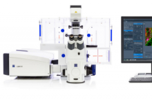
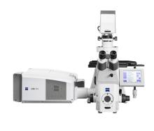
Airyscan2 LSM980 inverted confocal imaging system
For fast super-resolution, live cell imaging, we offer a fully automated Airyscan2 inverted confocal microscope. The Airyscan 2 allows for super resolution 3D imaging and 8x speed boost (compared to confocal at Nyquist), as well as tiled imaging, z-stacks and multi-position time-lapse imaging of living cells.
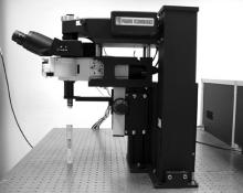
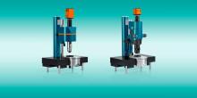
Prairie Ultima IV in vivo two-photon imaging system
For in vivo two-photon imaging, we offer a Prairie Ultima IV microscope equipped for isoflurane gas anesthesia and a temperature control system. This is one of two two-photon microscopes that share two Ti:sapphire lasers on a large optical table. (Note: Light box, faraday cage and XY stage not shown.)
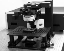
Prairie Ultima XY in vitro two-photon imaging system
For two-photon imaging of tissue slices, we offer a Prairie Ultima XY microscope equipped for whole-cell patch clamp recording, with an AxoPatch 1D amplifier and Sutter MP-285 computer-controlled micromanipulator. This is one of two two-photon microscopes that share two Ti:sapphire lasers on a large optical table. (NOTE: Light box and faraday cage not shown.)
Zeiss Axioimager fully automated DIC/fluorescence widefield microscope
The AxioImager is a fully-automated upright microscope equipped for both transmitted light (DIC) and fluorescence microscopy, allowing tiled images, z-stacks and time-lapse imaging.
Miltenyi / LaVision Biotec Ultramicroscope II
For light-sheet imaging of cleared tissue volumes, we offer a Miltenyi / LaVision Biotec Ultramiscroscope with six bi-directional light sheets for optimal even coverage of large tissue volumes (e.g., Whole Brain, Whole Heart, etc.)
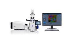
Zeiss LSM710 live cell imaging confocal microscope
The LSM 710 Confocal is a fully automated inverted laser-scanning confocal microscope equipped with an environmental control chamber, motorized stage and software allowing tiled images, z-stacks and time-lapse imaging of living cells.
Imaging Systems in our Lorry I. Lokey Stem Cell Building Facility
Airyscan2 LSM980 Plus NIR and Spectral Detection with Airyscan Joint Deconvolution
The new LSM980 provides live and fixed tissue imaging with super-resolution (up to 90nm) confocal capabilities coupled with near-IR excitation and detection with spectral scanning functionality.
Image processing workstations
Additionally, the NMS provides image processing workstations featuring Xeon Cores, 64GB+ RAM, and High End GPUs as well as advanced software resources for 3D, 4D interactive and volume visualization of data sets through python, Mathworks Matlab, FIJI, MBF Biosciences Neurolucida360, and Bitplane Imaris.