Image
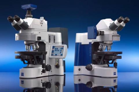
The AxioImager is a motorized, upright microscope equipped for both transmitted light (DIC) and fluorescence microscopy.
This microscope is perfect for Prof. Stephen Smith's Array Tomography technique, as well as general-purpose widefield microscopy.
Fast Facts
- Fully automated microscope allowing tiled images, z-stacks and time-lapse imaging
- Dual CCD cameras: monochrome and color
- Four-color multi-channel fluorescence with monochrome CCD
- Histological stains with color CCD camera.
Technical Specifications
| Z drive | DC motorized Z-drive with optoelectronic coding, resolution 10 nm, reproducibility ±10 nm |
|---|---|
| XY stage | Piezo-driven XY scanning stage with linear encoders, travel range: 225 × 85 mm, maximum speed: 100 mm/s, resolution: 0.2 μm, reproducibility: < 1 μm, absolute accuracy: ±5 μm |
| Transmitted light | 12 V/100 W halogen light source, manual condenser, 0.9 NA, brightfield/DIC/phase |
| Optovar | Manual 3-position tube lens: mag. 1.0×, mag. 1.6×, Bertrand lens |
| Fluorescence equipment | EXFO X-Cite 120 illumination source, coupled to scope with liquid light guide, motorized 6-position reflector turret with DIC cube and five fluorescence filter cubes (see below). |
| Camera | Zeiss AxioCam HRm Rev. 3. Full specifications below. |
| Software | Zen 2.3 with z-stack, automated tiling, autofocus and multi-positional imaging |
| Objectives (each with DIC slider) | ||||
|---|---|---|---|---|
| mag. | type | NA | working distance (mm) | pdf file |
| 2.5× | Plan Neofluar | 0.075 | 9.5 | click here |
| 10× | Plan Apochromat | 0.45 | 2.0 | click here |
| 20× | Plan Apochromat | 0.8 | 0.55 | click here |
| 40× | Plan Apochromat | 0.95 | 0.25 | click here |
| 63× (oil) | Plan Apochromat | 1.4 | 0.19 | click here |
| Fluorescence filter sets | |||||
|---|---|---|---|---|---|
| description | excitation peak/FWHM (nm) | dichroic 50% trans. (nm) | emission peak/FWHM (nm) | pdf file | |
| DAPI | 365/50 | 395 | 445/50 | DAPI.pdf | |
| eGFP | 470/40 | 495 | 525/50 | eGFP.pdf | |
| Cy3 | 550/25 | 570 | 605/70 | Cy3.pdf | |
| mCherry | 560/40 | 585 | 630/75 | mCherry.pdf | |
| Cy5 | 640/30 | 660 | 690/50 | Cy5.pdf | |
| Camera Specifications: AxioCam HRm Rev. 3 | ||||||||||||||
|---|---|---|---|---|---|---|---|---|---|---|---|---|---|---|
| Sensor | Sony ICX 285, progressive readout | |||||||||||||
| CCD Basic resolution | 1388 × 1040 = 1.4 megapixels | |||||||||||||
| Pixel size | 6.45 μm (h) × 6.45 μm (v) | |||||||||||||
| Sensor size | Chip area 8.9 mm × 6.7 mm (equivalent 2/3 inch) | |||||||||||||
| Spectral range | Approximately 350 nm–1000 nm with BK-7 protection glass without IR filter (BG40 can be inserted if necessary). | |||||||||||||
| NIR mode | Mode for higher sensitivity in near IR | |||||||||||||
| Dynamic range | Typical > 1:2200 at 25 MHz with < 7.7 e readout noise Typical > 1:2500 at 12.5 MHz with < 6.8 e readout noise | |||||||||||||
| Full well | Typical 17,000 e | |||||||||||||
| Dark current | Typical 0.7 e/pixel/s, optional dark current compensation for maximum low light performance at long exposure times | |||||||||||||
| Resolution improvement | Microscanning technology enables a configurable image resolution beyond the basic sensor resolution | |||||||||||||
| Selectable resolution | H × V (single shot) 276 × 208 (binning 5 × 5) 346 × 260 (binning 4 × 4) 462 × 346 (binning 3 × 3) 694 × 520 (binning 2 × 2) 1388 × 1040 (no binning) | H × V (microscanning) 2776 × 2080 (microscanning mode) 4164 × 3120 (microscanning mode) | ||||||||||||
| Live image frame rates |
| |||||||||||||
| Readout of sub-frames (ROI) | Random definition of regions of interest (ROI) on the sensor enables further increase of achievable frame rates | |||||||||||||
| Signal amplification | Analog: 2×, digital: 32× | |||||||||||||
| Digitization | Two switchable readout speeds offer different depths of digitization: High Quality: 14 bit / 12.5 MHz High Speed: 12 bit / 25 MHz | |||||||||||||
| CCD cooling | Single stage Peltier cooling, regulated | |||||||||||||
| Integration time | 1 ms up to several minutes | |||||||||||||
For an exploded diagram overview of this microscope, axioimagersystemoverview.pdf
For the complete AxioImager product brochure, zeissaxioimagerbrochure.pdf