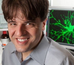
Optogenetics earns Stanford professor Karl Deisseroth the Keio prize in medicine
BY AMY ADAMS
Today optogenetics is a widely accepted technology for probing the inner workings of the brain, but a decade ago it was the source of some anxiety for then assistant professor of bioengineering Karl Deisseroth.
Deisseroth had sunk most of the funds he'd been given to start his lab at Stanford into a crazy idea – that with a little help from proteins found in pond scum he could turn neurons on and off in living animals, using light. If it didn't work he'd be out of funds with no published research, and likely looking for a new job.
Luckily, it worked, and has just earned Deisseroth, now the D. H. Chen Professor of bioengineering and of psychiatry and behavioral science, the 2014 Keio Medical Science Prize. Thousands of labs around the world are now using optogenetics to understand and develop treatments for diseases of the brain and mental health conditions and to better understand the complex wiring of our brains.
Deisseroth described the first step of his success in a seminal paper in 2005, but it was many years and many more academic papers before he could breathe easy. "There was a period of several years when not everyone who tried optogenetics got it working," Deisseroth said. "There were some people who were skeptical about how useful it would be, and rightly so because there were a number of problems we still had to solve."
Scientists worldwide have now used optogenetics to probe addiction, depression, Parkinson's disease, autism, pain, stroke and myriad other conditions.
"Optogenetics has revolutionized neuroscience," says Rob Malenka, a professor of psychiatry and behavioral sciences. Malenka is Deisseroth's former postdoctoral advisor and is now a frequent collaborator. "It has allowed neuroscientists to manipulate neural activity in a rigorous and sophisticated way and in a manner that was unimaginable 15-20 years ago."
Deisseroth adds, "I thought it would work but wasn't sure it would quite reach this point."
Lights on
Many years before Deisseroth began tinkering with optogenetics, Francis Crick – the Nobel Prize winner who co-identified the structure of DNA – had argued that neuroscience needed a tool to control one type of cell in the brain while leaving the others unaltered. Such a tool, he said, would give neuroscientists a way of turning particular groups of neurons on and off to learn more about how the brain functions.
Decades later, scientists around the world were still discussing possible ways of carrying out that vision, including some approaches using light. Deisseroth decided to build his lab's approach around proteins called microbial opsins that are found in single-celled organisms. He began with an opsin from green algae (aka pond scum) called channelrhodopsin, discovered by the German scientist Peter Hegemann.
This protein responds to light in a manner that is related to how neurons fire – it forms a channel on the cell surface that opens and allows ions into the cell. In the nerve, that channel opens when it receives a signal to fire. In the algae, it opens in response to light. If channelrhodopsin could be made to do the same thing in a neuron of a living animal it might provide a way of controlling the activity of that neuron using light – perhaps even during animal behaviors.
The concept seemed theoretically possible but risky, and indeed getting it to work in living animals took many years. Delivering the required large number of opsins into specific groups of cells within the brain, and targeting light deep into the brain, both posed challenges, and many opsins were initially not well-produced or tolerated by neurons.
When all the key pieces finally fell into place, they were able to use fiber optics to shine a tiny light onto a group of neurons harboring opsins in living animals and cause just that specific subset of neurons to either fire or be silenced in ways that controlled normal or disease-related behaviors.
"Karl had the insight to realize how important this was going to be," says Malenka, who is also the Nancy Friend Pritzker professor. "The idea had been floating around but he recognized the importance of the channelrhodopsin discovery and made it work."
Helping patients
Deisseroth believes the most important contribution from optogenetics will be its role in understanding biology and behavior, but says it will also likely contribute to medical advances.
One recent example of how optogenetics might directly help patients comes from a study by Gary Steinberg, professor of neurosurgery and neurosciences, who used optogenetics to activate a part of the brain in animals with a form of stroke and greatly improved their recovery.
Despite that promise, Deisseroth says he sees greater near-term potential for helping patients simply by applying lessons learned through optogenetics.
"There's no technical barrier to direct application," Deisseroth says, adding, "I'm risk-taking in my lab but quite different in the clinical setting."
Optogenetics involves genetically engineering neurons to produce foreign proteins, and once those proteins are in they can't come out. Deisseroth, Malenka and their colleagues Thomas Sudhof, a professor of medicine and Nobel prize winner, and Scott Delp, professor of bioengineering and of mechanical engineering, have started a company to overcome those barriers, but say that for now optogenetics is more likely to inform drug discovery or guide existing neural stimulation techniques.
One example of how optogenetics could point to better therapies came from work in Parkinson's disease. Deisseroth and his team used optogenetics to stimulate different components of the brain's wiring in animals with a version of Parkinson's disease, and found that connections arriving into a particular region deep in the brain, when stimulated, powerfully reduce symptoms.
Doctors who treat Parkinson's disease will sometimes implant an electrode that stimulates this brain region in patients, with quite a bit of success in reducing symptoms. But there had been debate over what wiring the electrode should stimulate. Deisseroth's work pointed to these arriving neuronal connections.
"Now neurosurgeons are finding that placing their electrical contacts to target connections gives better results in treating symptoms in people with Parkinson's and many other conditions," Deisseroth said.
Other examples have involved finding wiring that, when stimulated, reduces addiction, and similar lessons have been learned by Deisseroth and his collaborators about depression, anxiety, social behavior abnormalities and pain, among many other conditions.
Peripheral nerves
When people hear "neuron" they think "brain," but that overlooks the complex network of nerves running throughout our bodies that trigger muscles to move and that signal pain, touch and temperature to our brain.
Delp, who is also the James H. Clark Professor, has long had an interest in muscle movement and saw in optogenetics a possible way of triggering muscles to contract in people with movement disorders, such spinal cord injuries or spasticity from cerebral palsy.
Delp and students in his lab created a cuff that surrounds the nerve running down the back leg of mice. It stimulated the nerves that control leg muscles in a carefully orchestrated way that mimicked normal movement, and that didn't exhaust muscles the way other approaches had.
While working on muscle movement, the group found that they'd gotten opsins into the nerves that sense our environment. That kicked off a new line of research.
Graduate students Kathryn Montgomery and Shrivats Iyer followed up on that discovery, and got opsins into the nerves that control pain – a task that involved carefully injecting nerves the size of dental floss to insert the opsins. They were able to turn those nerves on and off using light, increasing or decreasing a mild sensation that the animals were freely able to move away from. Delp thinks this could be a powerful way of learning what causes chronic pain, and creating new therapies.
"This is an entirely new approach to study a huge public health issue," says Delp. "It's a completely new tool that is now available to neuroscientists everywhere."
Looking ahead
Much of the progress that has been made so far with optogenetics has come out of collaborations between Deisseroth and colleagues from the schools of Medicine and Engineering, and through students in his lab, 11 of whom have received Stanford Bio-X funding that allows them to share mentorship with other diverse faculty members.
The same interdisciplinary approach that got them to this point is driving future projects. Deisseroth says one important development comes out of advances in optics. Two students are now working on different approaches to guide light so precisely they can activate one nerve cell at a time.
"If you have a thousand cells in your field of view, you can say 'I want to turn on these 10 cells in a particular pattern during a behavior,'" Deisseroth said.
Another goal is to combine optogenetics with a different technique that came out of his lab, called CLARITY, which generates a three-dimensional view of the brain's connections. After carrying out optogenetics on a particular region of the brain, CLARITY would reveal precisely where those neurons connect throughout the brain.
For most studies optogenetic stimulation is delivered through a thin fiber-optic wire implanted into the mouse, which allows the scientists to modulate neurons deep in the brain. Members of Deisseroth's lab are working with Ada Poon, assistant professor of electrical engineering, who has expertise in wireless technology. They are developing a wireless way of delivering optogenetic stimulation.
"In the final version there will be a silicon chip with a transistor and controller and it will wirelessly connect with the source," Poon says. That is expected to be available within the next year, and could allow optogenetics to be combined with tiny microscopes to record the brain's activity.
Pond scum to the rescue?
Decades ago, when teams of scientists began studying the light-sensitive proteins within microbes it wasn't with an eye toward one day helping people with depression, Parkinson's disease or untreatable pain.
It was to better understand the intricate and amazing world around us. The research was fueled by pure scientific curiosity. Curiosity that has, as it happens, led to a discovery that might just help people.
Deisseroth argued in a Scientific American piece about the importance of funding curiosity-driven research as opposed to more targeted disease-focus research that some scientists and funding agencies have advocated.
"The more directed and targeted research becomes, the more likely we are to slow our progress, and the more certain it is that the distant and untraveled realms, where truly disruptive ideas can arise, will be utterly cut off from our common scientific journey," Deisseroth wrote.
He argues that funding agencies need to not only support fundamental research, but also facilitate the translation of that basic research into the work that can one day help patients.
"This is something that Bio-X does well," Deisseroth said. He works in the Clark Center that houses Bio-X, where scientists from very different backgrounds work elbow to elbow. "They put people who come from different perspectives within shouting distance of each other so those leaps can happen," he said. Bio-X has provided seed funding for a number of optogenetics collaborations and also supports the optogenetics core, where scientists can learn how to employ the technique in their own labs.
We need to be supporting people who are fascinated by pond scum and other obscure topics, Deisseroth argues, if we are to eventually treat depression, autism, Parkinson's disease and a host of other complex diseases.