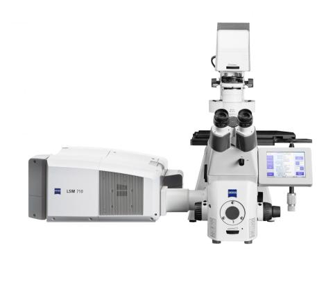
The LSM 710 Confocal is an inverted laser-scanning confocal microscope. This scope is equipped with an environmental control chamber, fully automated motorized stage and software for time-lapse imaging of living cells.
Fast Facts
- Fully automated microscope allowing tiled images, z-stacks and time-lapse imaging
- Temperature, humidity, and CO2 control for live imaging.
- Detection from 390 to 750 nm freely configurable
- Brightfield, DIC and polarizer
- Laser lines: 405nm; 488nm; 514nm; 568nm; 633 nm.
Specifications
AxioObserver.Z1 Microscope
| Z drive | DC motorized Z-drive with optoelectronic coding, resolution 10 nm, reproducibility ±10 nm. |
|---|---|
| XY stage | Motor-driven XY scanning stage (120 × 100 mm) with Mark & Find and Tile Scan (Mosaic Scan) functions; smallest increment 1 μm. |
| Transmitted light | 12 V/100 W halogen light source, motorized condenser LD 0.55 NA, brightfield/DIC/phase |
| Fluorescence equipment | EXFO X-Cite 120 illumination source, coupled to scope with liquid light guide, motorized 6-position reflector turret with DIC cube and three fluorescence filter cubes (see below). |
Objectives
| mag. | type | NA | w.d. (mm) | |
| 10× | Plan Apochromat | 0.45 | 2.0 | click here |
| 20× | Plan Apochromat | 0.8 | 0.55 | click here |
| 40× | Plan Neofluar (oil) | 1.3 | 0.21 | click here |
| 63× | Plan Apochromat (oil) | 1.4 | 0.19 | click here |
Fluorescence filter cubes
| Fluorescence filter cubes (note: no camera, these are for eyepiece viewing only) | ||||
|---|---|---|---|---|
| description | excitation | dichroic 50% trans. (nm) | emission | pdf file |
| DAPI | 365/60 (peak/FWHM, nm) | 395 | 420-470 (bandpass range, nm) | DAPI_set49.pdf |
| GFP | 450-490 (bandpass range, nm) | 490 | 500-550 (bandpass range, nm) | GFP_set38HE.pdf |
| DsRed | 545/25 (peak/FWHM) | 570 | 570-640 (bandpass range, nm) | DsRed_set43.pdf |
LSM 710 Scanning Module
| Scanner | Two independent galvanometric scanning mirrors, real-time controlled. |
|---|---|
| Scan resolution | 4 × 1 to 6144 × 6144 pixels, continuously variable |
| Scanning speed | 14 × 2 speed stages; up to 12.5 frames/s with 256 × 256 pixels; 5 frames/s with 512 × 512 pixels; (max. 77 frames/s with 512 × 32 pixels), 0.38 ms for a line of 512 pixels; up to 2619 lines per second. |
| Scan zoom | 0.6× to 40×, digitally variable in steps of 0.1 |
| Scan rotation | Free 360° rotation in steps of 1°, free X/Y offset. |
| Scan field | 20 mm diagonal field (max.) in the intermediate image plane, with full pupil illumination |
| Pinholes | Master pinhole pre-adjusted in size and position, individually variable for multi-tracking and short wavelengths (e.g. 405 nm) |
| Detection | Three simultaneous confocal fluorescence channels with highly sensitive low dark noise photomultiplier tubes (PMTs). Transmitted light channel with PMT. |
| Data depth | 8- 12- or 16-bit selectable |
Laser Module
| VIS Laser Module | Pigtail-coupled lasers with polarization-preserving single-mode fibers, temperature-stabilized VIS-AOTF (acousto-optical tunable filter) for simultaneous intensity control; switching time <5 μs |
|---|
| Laser | Line(s) (nm) | Power [end-of-life specification] (mW) |
| diode | 405 | 30 |
| Argon | 458, 488, 514 | 25 or 35 |
| DPSS (diode-pumped solid-state) | 561 | 20 |
| HeNe | 633 | 5 |
For a brief introduction to confocal_microscopy_from_Zeiss.pdf
