Stanford psychologists discover new patterns of brain development in areas linked to reading and face recognition
By Nathan Collins
As children learn to read and recognize new faces, connections in the brain associated with those tasks become better insulated and robust, Stanford researchers argue in a new study.
The research suggests a resolution to a debate about what happens in developing brains. Some measurements of brain regions associated with reading and face recognition indicated those regions were actually thinning out. In fact, Stanford psychologists argue September 23 in Proceedings of the National Academy of Sciences, that finding is an artifact associated with the growth of myelin, a tissue that insulates electrical connections between brain cells.
“These findings have important implications for understanding both typical and atypical brain development as they show that as the visual brain develops it likely becomes more efficient due to improved connections,” said Kalanit Grill-Spector, a professor of psychology and the new study’s senior author.
The mysterious shrinking brain
Although the new study focuses on the development of brain areas involved in reading and face recognition in school-aged children, its central question concerns a different, mystifying observation: as it develops, the top layer of the brain, known as the cortex, appears to thin by as much as a third. In other words, data from brain scans make it look a lot like kids are actually losing brain tissue as they learn to read, recognize their friends and family, and so on.
The dominant explanation, said Grill-Spector, who is also a member of Stanford Bio-X and the Wu Tsai Neurosciences Institute, is pruning. Very early in development, during infancy, neuroscientists know, the brain has an excess of connections. In order to make brain circuits more specialized, the brain removes or prunes, unnecessary connections. Among other things, pruning occurs in infancy between neurons in our brains’ primary vision processing circuits, so it was plausible that pruning might continue in school-aged children and support other visual circuits involved in reading and face recognition.
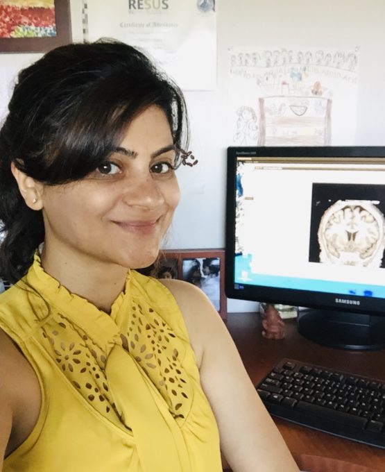
 Vaidehi Natu, a postdoctoral fellow in psychology, led the study. (Image credit: Courtesy Vaidehi Natu)
But Grill-Spector, her postdoctoral fellow Vaidehi Natu, and colleagues also considered another possibility: Perhaps the cortex isn’t shrinking as much as we thought. Instead, it might be that as children grow, their brains might contain more of a material called myelin, which shields the fibers that connect brain cells together and improves signal transmission between them. That increase in myelin is a good thing since it improves signal transmission between neurons, but it poses a problem for measuring cortex thickness when using standard magnetic resonance imaging, or MRI, brain scans.
“You can imagine the cortex to be like pavement and the myelinated fibers like grass growing beside the pavement,” Natu said. “As the grass grows and covers the pavement, it is harder to see its edge.” In the same way, myelin can obscure the boundary between cortex and adjacent tissue containing neural fibers, making it hard to measure cortex thickness.
Inside kids’ brains
To test that idea, the team scanned the brains of 27 kids 5 to 12 years old along with 30 grownups, focusing on regions of the brain related to word, face and place recognition. The team ran three kinds of magnetic resonance imaging brain scans, including two newer techniques that are more sensitive to myelin.
As they had suspected, they found tissue growth that was associated with myelin in children’s brains. But the tissue growth related to myelin wasn’t everywhere. While the amount of myelin seemed to grow in parts of the brain associated with word and face recognition, it did not grow in areas associated with recognizing places. That’s consistent with the fact that brain regions involved in face and word recognition develop over a longer period during childhood and adolescence compared to the region involved in recognizing places, Grill-Spector said.
In collaboration with Evgeniya Kirilina, a researcher at the Max Planck Institute of Human and Cognitive Brain Sciences, the team confirmed differences in myelination across brain regions in post-mortem analyses of adult brains.
“When we first saw myelin in the cortex in the images obtained by the microscope, we were struck, because you don’t get to see that usually,” Natu said.
With support from a Big Ideas grant, Grill-Spector, Jennifer McNab, a research associate professor of radiology, and Daniel Yamins, an assistant professor of psychology will begin to address those issues. The team will use brain-scanning technologies, tissue analysis and computer science to try to understand how infant brains support learning and change in response to it.
“We know even less about the infant brain, because it’s so challenging to obtain data,” Grill-Spector said. “It’s going to be groundbreaking and interesting.”
Additional authors include Jesse Gomez, a recent graduate of the Stanford Neurosciences PhD Program, and researchers from Max Planck Institute for Human and Cognitive Brain Sciences, Leipzig Germany, and the University of California, Berkeley.
The research was funded by grants from the National Institutes of Health, a National Research Service Award, the European Research Council and the German Federal Ministry of Education and Research.
To read all stories about Stanford science, subscribe to the biweekly Stanford Science Digest.
Media Contacts
Nathan Collins, Stanford News Service: (650) 725-9364, nac@stanford.edu
