It’s time to revamp the motor homunculus

For almost a century, budding neuroscientists have been taught that the headband-like strip of brain tissue over our ears that controls our movements, called the motor cortex, contains an orderly map of our bodies. Brain cells concerned with moving each body part – from the tips of our toes to the tips of our fingers – are all laid out in sequence, as well as a large zone dedicated to our fabulously expressive faces and speech muscles. That’s the textbook account, anyway.
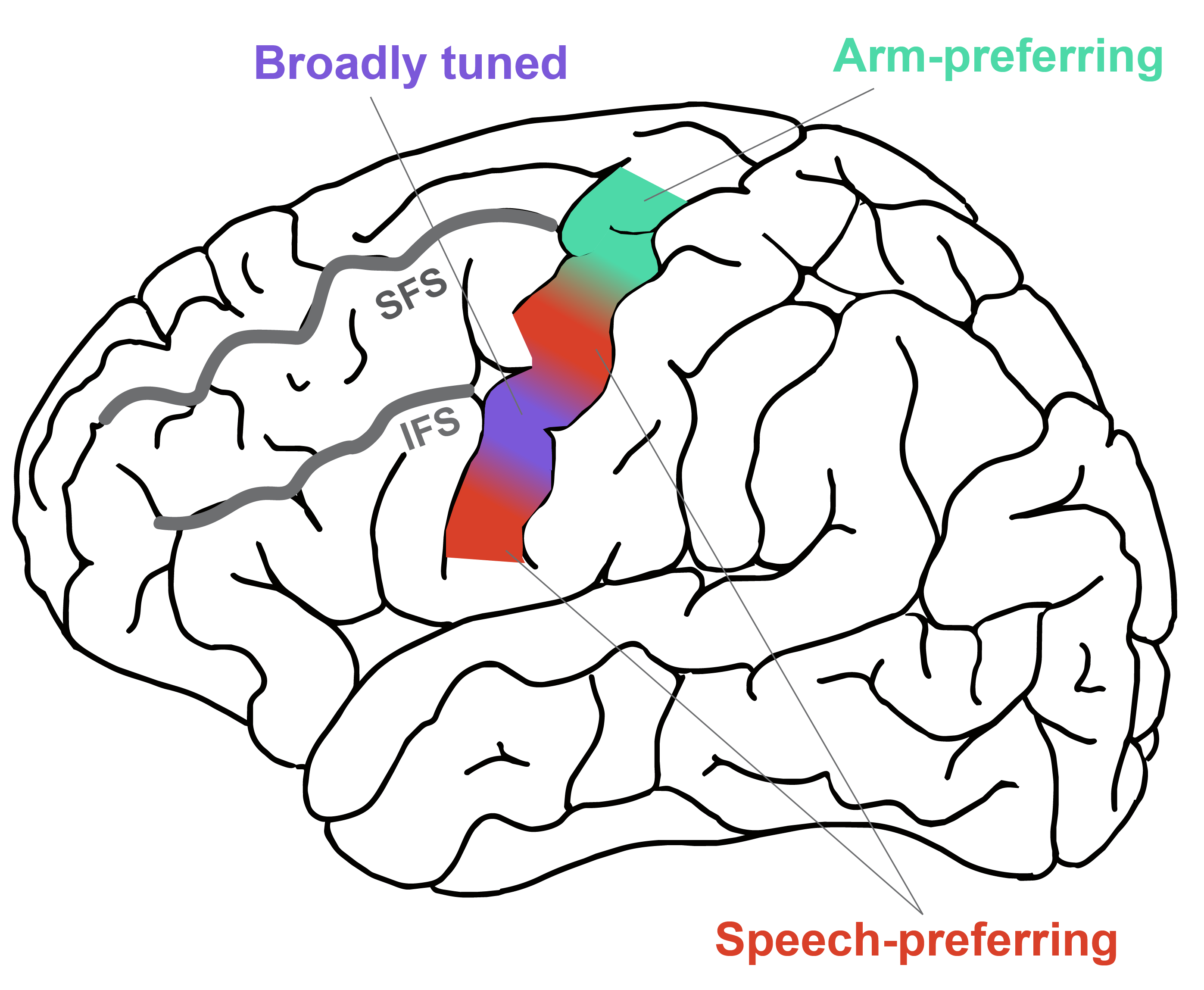
But that extremely satisfying model, known as the motor homunculus, doesn’t tell the full story, according to a new study by Wu Tsai Neurosciences Institute researchers, published June 17, 2026 in the journal Nature. The new study finds that many parts of the motor cortex have information about the entire body. The region that is predominantly associated with movement of the arms, for example, also has some information about movements of the face, and the face region has some information about movement of the legs.
“It was quite exciting to see these whole-body representations appear,” said Darrel Deo, an instructor in the Department of Neurosurgery at Stanford Medicine and the new paper’s first author.
Apart from a deeper understanding of the motor cortex, the results could also lead to better brain-computer interfaces (BCIs) – systems that translate brain activity into signals that control or interact with devices that aid people who’ve lost the ability to move or communicate because of neurological disease or paralysis. The new study suggests a single implant could provide access to a wider repertoire of movement-related control signals than previously appreciated, Deo said, rather than one implant for each affected region of their body.
Deo began the work as a Wu Tsai Neuro postdoctoral scholar in the lab of the late Krishna Shenoy and Jaimie Henderson, the John and Jene Blue - Robert and Ruth Halperin Professor and a professor of neurosurgery at Stanford Medicine. As part of that effort, Deo and lab co-director Frank Willett, an assistant professor of neurosurgery at Stanford Medicine, wanted to update the motor homunculus map with modern tools. “We were keen to revisit the motor cortex at a much finer scale,” Deo said.
To do so, Deo and his colleagues studied eight participants who were already enrolled in ongoing BCI clinical trials – six from the BrainGate2 clinical trial and two from a separate clinical trial run through the University of Pittsburgh and University of Chicago. These participants were all implanted with microelectrode arrays capable of recording electrical signals from individual neurons in various locations along their motor cortex – something that wasn’t possible when the motor homunculus idea was first developed.
Then, Deo asked the participants to perform 45 different body movements, such as lifting a foot or turning a hand (or to attempt the movement if they were no longer able to move that body part) as he recorded activity from motor cortex neurons.
In each and every region the researchers looked at, Deo said, “there was distinguishable information about movements that span the entire body, not just the canonical body part the motor homunculus model would have suggested.” In other words, while one region might be most strongly activated for hand movements, it also contains signals concerning movements in the face, legs, and everywhere else.
The researchers also propose a new, modified map of the motor cortex that differs from the original homunculus model. In this new map, two areas tuned for speech flank the superior ventral precentral gyrus, which the team found was broadly tuned to all parts of the body. “It’s like a jack-of-all-trades area,” Deo said.
Those results could be a boon for BCI design. For example, a single electrode array implanted in the superior ventral precentral gyrus could be used to control robotic arms, communication devices, and more.
More generally, the study suggests it’s time to move beyond the motor homunculus’s strict partitioning of the motor cortex. “It’s a bit more intermixed than we thought,” Deo said.
Publication details
Research Team
Study authors were Darrel Deo from the Department of Neurosurgery at Stanford Medicine and the Wu Tsai Neurosciences Institute; Elizaveta V. Okorokova from the University of Chicago and the University of California, Davis; Anna L. Pritchard, Samuel R. Nason-Tomaszewski, Deqiang Qiu, Nicholas AuYong, and Chethan Pandarinath from the Georgia Tech University and Emory University; Nick V. Hahn, Eun Young Choi, Foram B. Kamdar from the Department of Neurosurgery at Stanford Medicine; Nicholas S. Card, Maitreyee Wairagkar, Carrina Iacobacci, David M. Brandman, and Sergey D. Stavisky from the University of California, Davis; Justin Jude, Claire Nicolas, Alexander Acosta, Sydney S. Cash, Daniel B. Rubin, and Ziv M. Williams from Harvard Medical School and Massachusetts General Hospital; Thomas Hosman from Providence VA Medical Center and Brown University; Yuguang Meng from Emory University; Leigh R. Hochberg from Harvard Medical School, Providence VA Medical Center, and Brown University; John E. Downey and Sliman J. Bensmaia from the University of Chicago; Jaimie M. Henderson from the Department of Neurosurgery at Stanford Medicine, the Wu Tsai Neurosciences Institute, and Stanford Bio-X; and Francis R. Willett from the Department of Neurosurgery at Stanford Medicine.
Research Funding
This work was supported by the NIH National Institute of Neurological Disorders and Stroke (U01NS123101 and UH3NS107714); the NIH National Institute on Deafness and Other Communication Disorders (R01DC014034 and K23DC021297); the Wu Tsai Neurosciences Institute; the Howard Hughes Medical Institute; Larry and Pamela Garlick; the Office of Research and Development, Rehabilitation R&D Service, US Department of Veterans Affairs (A2295R and N2864C); the AHA’s Second Century Early Faculty Independence Award; and the Office of the Assistant Secretary of Defense for Health Affairs through the Amyotrophic Lateral Sclerosis Research Program under award number AL220043.
Competing Interests
The MGH Translational Research Center has a clinical research support agreement with Axoft, Neuralink, Neurobionics, Precision Neuro, Synchron and Reach Neuro, for which Hochberg provides consultative input. Hochberg is a co-investigator on an NIH SBIR grant with Paradromics, and is a non-compensated member of the Board of Directors of a non-profit assistive communication device technology foundation (Speak Your Mind Foundation). Mass General Brigham is convening the Implantable Brain-Computer Interface Collaborative Community (iBCI-CC); charitable gift agreements to Mass General Brigham, including those received to date from Paradromics, Synchron, Precision Neuro, Neuralink and Blackrock Neurotech, support the iBCI-CC, for which Hochberg provides effort. Henderson is a consultant for Paradromics; serves on the Medical Advisory Board of Enspire DBS; is a shareholder in Maplight Therapeutics; and is an inventor on intellectual property licensed by Stanford University to Blackrock Neurotech and Neuralink. Stavisky is an inventor on intellectual property licensed by Stanford University to Blackrock Neurotech and Neuralink; and is currently an advisor to Sonera. Frank Willet is an inventor on intellectual property licensed by Stanford University to Blackrock Neurotech and Neuralink. Choi is a consultant for Neuralink. Pandarinath is a consultant for Meta (Reality Labs) and Synchron. D.M.B. was a surgical consultant for Paradromics. Stavisky, Brandman, Card, and Wairagkar are inventors of intellectual property related to neuroprostheses owned by the University of California, Davis. All other authors declare no competing interests.


