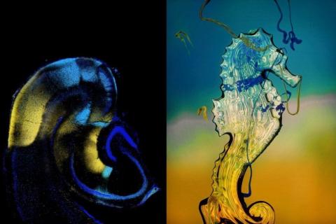
Attractive and repulsive forces between two multitasking molecules help assemble neural circuits in mice, Stanford study finds
Holly Alyssa MacCormick
Multitasking molecules may be the key to solving the riddle of how the brain makes trillions of specifically coded connections between the brain cells known as neurons.
In mice, two molecules that stud cell surfaces can create attractive or repulsive forces between the brain cells they’re displayed on, a new Stanford University study has found. By attracting or repelling other brain cells questing for neural connections, the magnet-like molecules called teneurin-3 and latrophilin-2 play an important role in wiring the part of the brain that helps mice navigate and process information about the objects around them.
The findings help explain how multitudes of neurons can connect in an ordered way using relatively few molecular “tags,” according to a study led by Stanford biologist Liqun Luo, the Ann and Bill Swindells Professor in Stanford’s School of Humanities and Sciences, and Daniel Pederick, lead author and postdoctoral research fellow in the Department of Biology. The study also reveals the architecture of brain development in one segment of the hippocampus, the part of the brain that encodes, stores, and retrieves memories. The research was published June 4 in the journal Science.
A wiring problem
One of the brain’s many mysteries is how it’s wired. As the brain grows, neural networks form when neurons connect to one another via threadlike projections, called axons. The exact order of neural assembly is unknown, but the brain’s connections are thought to be individually coded using a kind of tagging system, and cell surface molecules are known to act as cues for axons seeking partners.
But herein lies a conundrum: “The human brain contains about 100 billion neurons making more than 100 trillion connections. But the human genome has only 20,000 genes, and only about 1,000 to 2,000 of them are expressed on the cell surface and can serve as the cell surface tagging molecules for brain wiring,” explained Luo, the study’s senior author. “The question is, how can a small number of molecules help so many neurons make so many connections so precisely?”
Previous research by Luo’s lab provided some clues. Research published in the journal Nature in 2012 by former graduate student Weizhe Hong revealed that a family of cell surface molecules, called teneurins, can play a role in partner matching for neurons in fruit flies. Nic Berns, another former graduate student in Luo’s lab, showed how neurons in two different parts of the hippocampus could find each other simply by looking for other neurons that display the same molecule (teneurin-3) on their cell surfaces. This research was published in the journal Nature in 2018.
“We wanted to find a cell surface molecule that was present in an opposite pattern to teneurin-3 during hippocampal development,” explained Pederick. The researchers thought that such a molecule could be involved in neural network assembly and may shed light on how the two parallel networks in that brain region form together.
Parallel neural networks are important because they enable the brain to simultaneously process more than one type of information. In the region of the brain that Pederick, Luo and their colleagues studied, parallel networks simultaneously process memories related to spatial and object-related information, such as recalling where you parked your car and what it looks like.
Pederick worked with Luo lab research scientist Jan Lui to search for a cell surface molecule that was present in the opposite places teneurin-3 was found in the two parallel networks in the mouse hippocampus.
The researchers examined brain cells from the mouse hippocampus using a technique that sorts individual cells according to their unique properties. This work was done in the in the lab of Stephen Quake, the Lee Otterson professor in the School of Bioengineering. This technique, called fluorescence-activated cell sorting-based single-cell RNA sequencing, helped the researchers identify latrophilin-2 as their molecule of interest.
However, the role of latrophilin-2 in neural network assembly was not yet known. “It was another level of intrigue,” Pederick said.
Making genes disappear
The researchers performed genetic manipulations to investigate the roles of latrophilin-2 and teneurin-3 in mouse hippocampus wiring. These genetic experiments removed latrophilin-2 from certain parts of the mouse hippocampus, or added it to areas where it didn’t naturally exist, to test how it might change the assembly of neural circuits.
The scientists predicted that latrophilin-2 was a cue that would repel axons expressing teneurin-3 on their cell surfaces given their opposite expression patterns.
“We knew the area of the hippocampus where axons expressing the cell surface molecule teneurin-3 would normally go, so we deliberately overexpressed latrophilin-2 in small areas of that region,” Pederick explained. “In the same brain, we left unaltered, normal areas.”
By creating a mosaic of unaltered and latrophilin-2 rich patches in living mouse brains, the researchers were able to do side-by-side comparisons to observe the possible effects of latrophilin-2 on axons that had teneurin-3 on their cell surfaces.
The results confirmed their prediction, but with an additional discovery.
In the medial neural network of the mouse hippocampus, teneurin-3-expressing axons were attracted to teneurin-3 targets and were repelled by latrophilin-2 targets, as the researchers predicted. But when the researchers tested the reverse configuration in the lateral network, they found latrophilin-2-expressing axons were repelled from teneurin-3 targets.
The versatility of these molecules was unexpected. “We identified this pair of molecules, called teneurin-3 and latrophilin-2, that can serve as either a ligand (signal-sending molecule) or a receptor (receiving molecule) by attracting and repelling axons, expanding the known functions of what each individual molecule can do,” Luo said.
One of the big findings in the study is the way the two parallel networks are separated in the mouse hippocampus, Pederick explained. “Instead of the networks each having their own attractive forces, there's attraction happening in one network, and repulsion happening between them.”
“The number of times the same molecules are used to ensure that these two networks develop separately was unexpected,” Pederick said.
This research explored molecular interactions in one part of the mouse hippocampus, but teneurin-3 and latrophilin-2 are found in similar ratios elsewhere in the hippocampus, suggesting the study’s results may apply more broadly, Luo explained.
The hippocampus encodes memories, and this foundational research expands our understanding of how this critical part of the brain is wired to work properly. Studies like this are also essential for researchers studying neural diseases that make memory go awry, the researchers explained.
“It's a beautiful story that solves a developmental neurobiology wiring problem in an elegant way,” said Luo.
Liqun Luo is also an investigator of the Howard Hughes Medical Institute, a professor (by courtesy) of Neurobiology, a member of Stanford Bio-X, a member of the Stanford Cancer Institute, a faculty fellow at Stanford ChEM-H, a member of the Wu Tsai Neurosciences Institute. Stanford co-authors Daniel Pederick, Jan Lui, Ellen Gingrich, Chuanyun Xu, Mark J. Wagner, and Liqun Luo are all members of the Department of Biology. Stephen Quake is a professor of Bioengineering, of Applied Physics and (by courtesy) of Physics. Quake is also co-president of the Chan Zuckerberg Biohub.
Co-authors Yuanyuan Liu and Zhigang He are members of the F.M. Kirby Neurobiology Center, Department of Neurology, Boston Children’s Hospital, Harvard Medical School. Liu is currently at the National Center for Complementary and Integrative Health at the National Institutes of Health.
This research was mainly supported by the National Institutes of Health and the Howard Hughes Medical Institute. Portions of the research were supported by the Neuro-Omics Initiative of the Big Idea Programs from Wu Tsai Neurosciences Institute at Stanford. Pederick was supported by an American Australian Association Education Fund Scholarship.
