Finalists announced for the 2016 Art of Neuroscience competition

Eleven images representing a broad cross section of neuroscience research have been chosen as finalists in the Stanford Neuroscience Institute’s Art of Neuroscience competition.
The images range from the intricately detailed, such as a spider-like neurons or arrays of cells that send visual signals to the brain, to the abstract, with a pen and ink illustration of neurons snuggled into a cozy home in the brain.
The images show both the beauty and the diversity of neuroscience research. Some were taken while probing the inner workings of neurons, while others depict brain-wide connections or the behavior of ants, which mimic our own brain networks. Two images show how stem cells are being used to understand brain development in a lab dish or for possible therapeutic uses.
Taken together, the images span the three key focus areas within the Stanford Neurosciences institute – NeuroDiscovery, NeuroEngineering and NeuroHealth – and represent the many research areas focused on healing and understanding our brains, and through that ourselves.
1st Place
Title of Image: Synaptogenesis
Description of image: Cultured hippocampal neurons were transfected with EGFP, and stained with a synapse marker, synapsin I and a dendrite/spine marker, ICAM-5. Synapses are formed opposed to the soma and dendrites.
Area of focus: NeuroDiscovery
Submitted by: Lin Ning, Postdoctoral Research Fellow, Neurobiology
Lab: Michael Lin, Assistant Professor of Neurobiology, Bioengineering and, by courtesy, of Chemical and Systems Biology
2nd Place
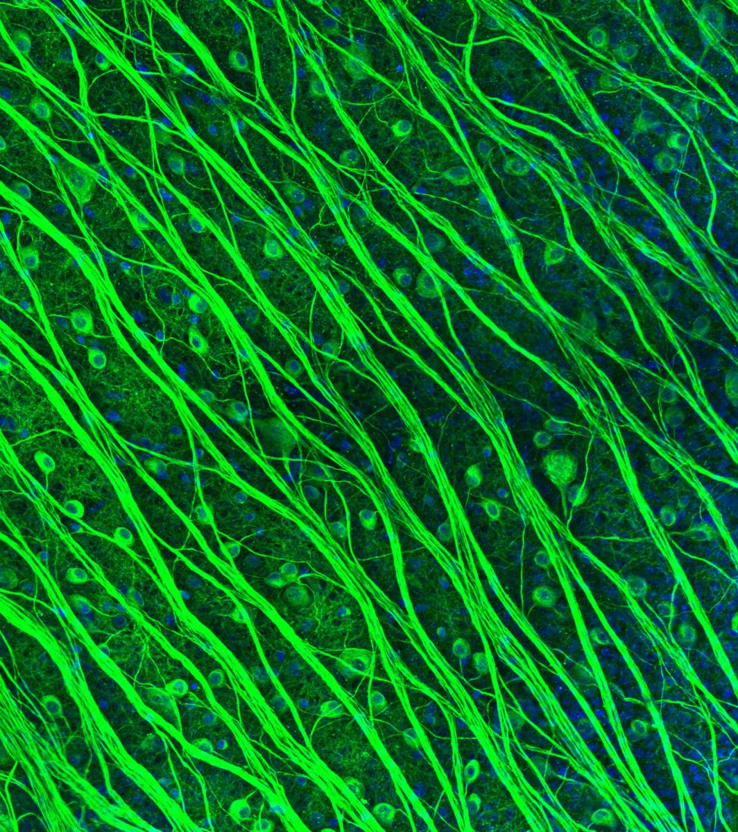
Title of Image: Retinal ganglion cells in the primate retina
Description of image: Immunostaining for ßIII tubulin in the primate retina reveals retinal ganglion cells (RGCs). RGC axons are arranged in bundles that carry visual signals through the optic nerve to the brain. Better understanding cells of the retina will allow retinal prostheses in development to tap into these cells and relay visual information to the brain.
Area of focus: NeuroEngineering
Submitted by: Lauren Grosberg, Postdoctoral Research Fellow, Neurosurgery
Lab: E.J. Chichilnisky, John R. Adler Professor, Professor of Neurosurgery and Ophthalmology and by courtesy of Electrical Engineering
Confocal image taken at the Neuroscience Microscopy Service
3rd Place
Title of Image: Hippocampus
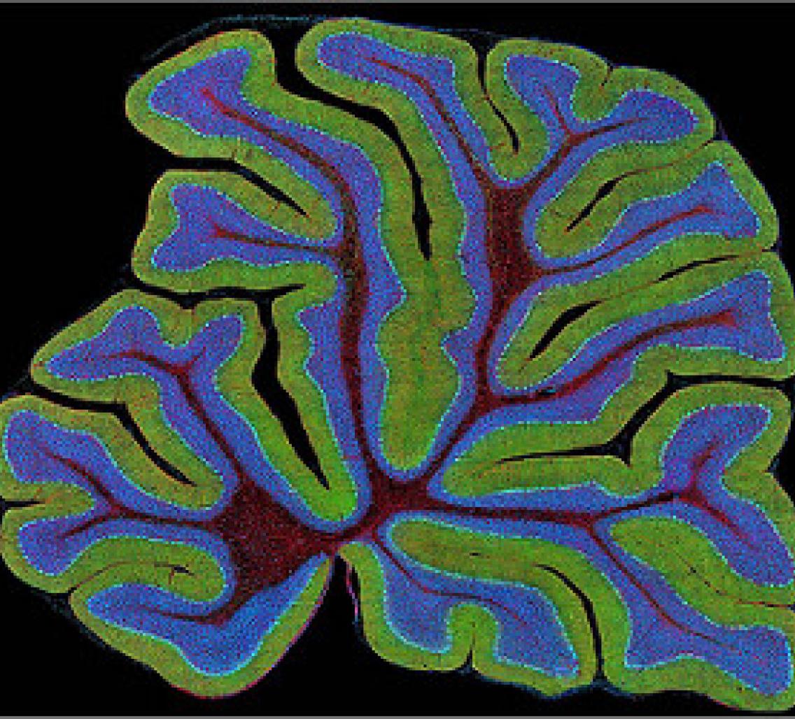
Description of Image: Golgi-stained hippocampus of a mouse model of Down syndrome
Area of focus: NeuroHealth
Submitted by: Ahmad Salehi, Clinical Professor, Psychiatry VA Research
Lab: Ahmad Salehi, Clinical Professor, Psychiatry VA Research
Honorable Mentions
Title of Image: Deception
Description of Image: Until recently, thymidine analogs which are taken up by dividing cells and incorporated into their DNA, were used to label stem cells transplanted into the brain in animal studies. Many types of cells, including those from bone marrow were claimed using this technique to be able to generate neurons. Unfortunately, this technique was later found to be unreliable, since many cells die after transplantation and the BrdU is then taken up by neighboring dividing cells that then masquerade as the transplanted cells. This image is from the study that revealed the flaws in the BrdU labeling technique. BrdU labeled cells were intentionally killed and transplanted into the brain of an adult mouse. This image demonstrates the BrdU that was taken up by dividing cells in the neurogenic subventricular zone of the recipient mouse brain. 50um coronal floating sections of formaldehyde-fixed brain were processed for immunofluorescence using antibodies against BrdU (red), GFAP (blue) and nestin (green).
Area of focus: NeuroDiscovery
Submitted by: Terry Burns, Neurosurgery Resident
Lab: Theo Palmer, Associate Professor of Neurosurgery
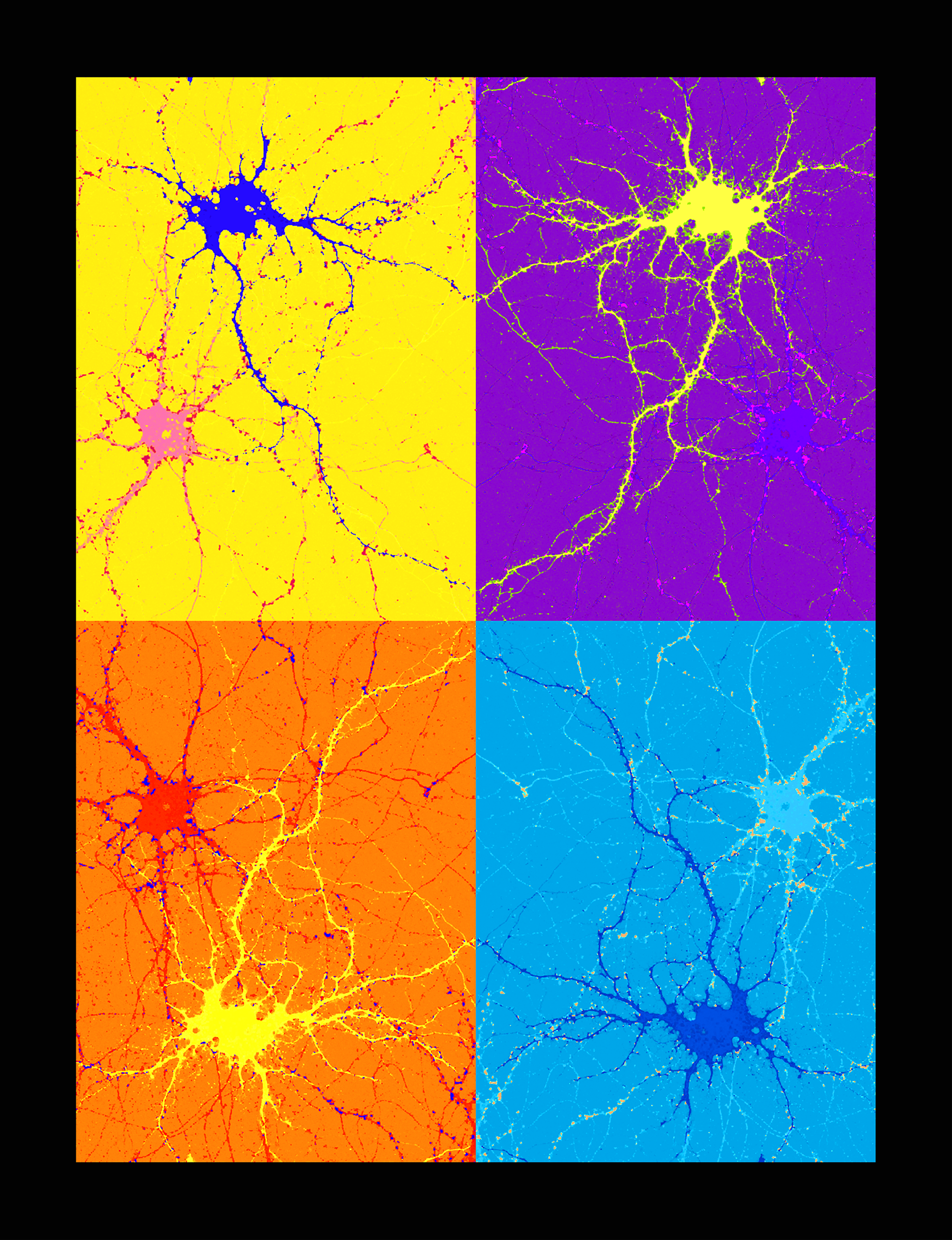
Title of Image: Mind's Labyrinth
Description of Image: Cultured mouse cortical neurons that have been stained using antibodies against a Major Histocompatibility Complex protein (Green) and synapsin (Red), a marker of presynaptic neural connections.
Area of focus: NeuroDiscovery
Submitted by: Kylie Chew, Postdoctoral Research Fellow, Biology
Lab: Carla Shatz, Sapp Family Provostial Professor, David Starr Jordan Director, Stanford BIO-X and Professor of Biology and Neurobiology
Title of Image: Colony interaction network
Description of Image: Brain networks operate by the combined actions of neurons, and an ant colony operates by the combined actions of many individual ants. We are investigating how ants use an evidence accumulation process to make foraging decisions. Films from field observations were used to track ants to obtain the paths shown here. The dots are locations of interactions between ants, and the colors represent different start and end locations for the trajectories. We constructed images similar to what is shown here to visualize how returning foragers (green and yellow) exchange information with potential foragers (blue and red), and how this influences ant foraging decisions. Although the ants only exchange local information by meeting other ants, these interactions form a colony-level interaction network and lead to the remarkable collective behavior response of the colony.
Area of focus: NeuroDiscovery
Submitted by: Jacob Davidson, Affiliate
Lab: Deborah Gordon, Professor of Biology
Title of Image: Oxytocin (Enter the Cuddle)
Description of Image: This is a freehand ink drawing of two oxytocinergic neurons snuggling amidst a sea of glia. Relationships - between humans or neurons - are defined by their context. And glia are as cuddly as can be.
Area of focus: NeuroDiscovery
Submitted by: Daniel Friedman, PH.D. student in Biology
Other Tech - Graduate, Daper Department wide Operations
Lab: Deborah Gordon, Professor of Biology
Title of Image: Mouse somatostatin-expressing hippocampal neuron
Description of Image: The somatostatin-expressing interneuron in mouse CA1 stratum oriens was recorded in whole-cell mode to reveal spiking and synaptic activity during intrinsically generated theta rhythm in an intact hippocampal preparation in vitro. Somatostatin+ str. oriens neurons displayed large heterogeneity in phase-locking to theta. Recorded neurons were labelled with neurobiotin and revealed with green fluorescent streptavidin. Somatostatin+ neurons were identified using tdTomato (red) and sections were reacted with Hoechst-33258 for nuclear staining (blue).
Area of focus: NeuroDiscovery
Submitted by: Carey Huh, Postdoctoral Scholar, Comparative Medicine
Lab: Shaul Hestrin, Professor of Comparative Medicine
Title of Image: Golden Gate Bridge
Description of Image: The structural connectivity of nerve fiber pathways measured with diffusion MRI in a coronal slice of the human brain. Tissue structures such as cell membranes restrict the self-diffusion of water and lead to anisotropic water diffusion along nerve fiber pathways. By observing the self-diffusion patterns of water during a diffusion MRI experiment we can reconstruct fiber pathways and get a view into the structural connectionsbetween different brain regions within the living brain. The colors in the image represent the fiber directions (blue: superior-inferior; green: anterior-posterior; red: left-right).
Area of focus: NeuroDiscovery
Submitted by: Christoph Leuze, SNI Interdisciplinary Scholar, Postdoctoral Research Fellow, Radiological Sciences
Lab: Jennifer McNab, Assistant Professor (Research) of Radiology (General Radiology)
Title of Image: Nerve fibers, blood vessels, and ganglion cell architecture of the retina
Description of Image: Ganglion cell layer of the primate retina, labeled with DAPI (red, neuron and blood vessel nuclei), beta-III Tubulin (blue, neurons and nerve fibers), and Ankyrin-G (green, axon initial segments). From the paper /Anatomical Identification of Extracellularly Recorded Cells in Large-Scale Multielectrode Recordings/ http://dx.doi.org/10.1523/JNEUROSCI.3675-14.2015. Better understanding cells of the retina will allow retinal prostheses in development to tap into these cells and relay visual information to the brain.
Area of focus: NeuroEngineering
Submitted by: Peter Li, Postdoctoral Research Fellow, Neurosurgery
Lab: E.J. Chichilnisky, John R. Adler Professor, Professor of Neurosurgery and Ophthalmology and by courtesy of Electrical Engineering
Title of Image: The blooming human cerebral cortex in a dish
Description of Image: Section of a human cortical spheroid derived from stem cells shows progenitors (pink) and dividing cells (green) around a ventricle-like structure. This 3D culture resembles the fetal cerebral cortex.
Area of focus: NeuroDiscovery
Submitted by: Anca Pasca, Postdoctoral Research Fellow, Neonatal and Developmental Medicine
Lab: Sergiu Pasca, Assistant Professor of Psychiatry and Behavioral Sciences - Stanford Center for Sleep Sciences and Medicine


