Q&A: ‘To see is to believe’
Imagine what researchers could do if they could see—literally see—all the neural activity going on deep inside a living animal’s brain. Along with light-based techniques to visualize brain activity, that level of access could radically transform what we know about the brain and perhaps pave the way for better treatments for disease.
But how would that be possible? The brain is opaque, and right now, seeing beyond the surface requires invasive surgery and implants. Except in rare cases involving naturally transparent animals, seeing activity unfold across a whole brain all at once is impossible.
Unless, that is, you figured out how to follow nature’s lead and made the brain see-through.
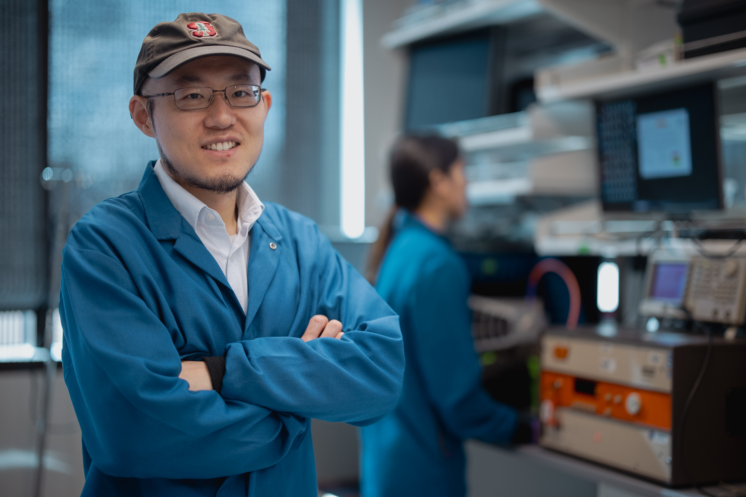
To Guosong Hong, the idea isn’t so far-fetched. As Wu Tsai Neurosciences Institute Faculty Scholar, he brings ideas from chemistry, physics, and engineering to multidisciplinary collaborations that address a key question: How can we better see what the brain is doing?
Early in his career, Hong—an assistant professor of materials science and engineering at Stanford’s School of Engineering—developed fluorescent dyes that could be lit up with infrared light. When injected inside animals, the dyes revealed previously unseen details of the brain and other tissues. Hong and his lab have also engineered materials for neural implants and methods to modulate neural activity. Most recently, he made the news for making mouse skin transparent with a common food dye, an advance that has already yielded new insights into brain development.
Now, a Big Ideas in Neuroscience grant from Wu Tsai Neuro will bring him together with biologists Lauren O’Connell and Xiaoke Chen, institute affiliates based in the Stanford School of Humanities and Sciences, to tackle what he calls “an even crazier idea”: genetically engineering a mouse with a transparent brain. The trio will study naturally clear glass frogs to understand how certain proteins make their bellies transparent, then create mice that produce similar proteins in their brains. That would make it possible to use methods like fluorescence imaging to track neural activity simultaneously across the entire brain.
“We are working across interdisciplinary boundaries. It’s like solving a puzzle from different angles. If one corner can’t be solved immediately, maybe you can come from a different angle and work on that part first. Eventually that’s going to lead to completing the entire puzzle.”
Guosong Hong
Such ideas are positioning Hong as an emerging voice at the interface of engineering and neuroscience. He was recently named a 2026 Sloan Research Fellow, and he was just awarded a prestigious MIND Prize from the Pershing Square Foundation, which will support him and his team as they draw on their discoveries to create an unprecedented map of neurodegeneration across the entire brain over time. That map could help researchers develop new insights into—and perhaps new treatments for—Alzheimer’s and other neurodegenerative diseases.
On the heels of those awards, we spoke to Hong to learn about his intellectual journey and mind-bending array of research projects.
How did you get interested in neuroscience?
I was trained as a physical chemist, and I had the habit of seeing the world through the lens of physics, focused primarily on the external world. That thinking changed during my fourth year of graduate school, when I read about President Obama’s BRAIN Initiative announcement. He said, “As humans, we can identify galaxies light-years away. We can study particles smaller than an atom. But we still haven’t unlocked the mystery of the three pounds of matter that sits between our ears.”
I realized that physics and chemistry principles, which have been incredibly successful at helping us understand the external world, could be turned inward. The brain is also a physical system. That realization set me on this journey into neuroscience.
What drew you toward the idea of making tissue transparent?
If we can get light deep in the tissue, we can better study its structure and function. Making the whole brain transparent has two advantages. It allows optogenetics, which lets us manipulate neural activity with light, and optical neural imaging, such as calcium and voltage imaging, to more easily target deep brain regions without implanted fibers or microendoscopes. And since the brain is a highly integrated system, the ability to image activity across the brain paints a more complete picture, which will help neuroscientists understand its many functions.
But it is notoriously difficult to get light into the brain. We have a lot of water in our brain, but it’s also lipid-rich. Light travels through water and lipids at different speeds, which causes the light to scatter and makes the tissue opaque.
I was reading a physics textbook and realized there was this very interesting physical principle from the 1920s called the Kramers-Kronig relations that seemed to be hidden in plain sight for biology research.
The idea is, if you make a material absorb more light at one wavelength, you can slow down light at other wavelengths. So if we apply a light-absorbing molecule to tissue, we can make light travel through water and lipids at similar speeds. That means the light will not scatter as much, and the tissue will become transparent.
Your team applied that idea to make a mouse’s skin transparent. How did that lead to your Big Ideas in Neuroscience project to create a mouse with a transparent brain?
After that paper, I had a discussion with Lauren O’Connell. She’s a biologist here at Stanford, and she studies glass frogs, which look transparent to the naked eye.
I thought this must be coming from the same principle. Glass frogs are transparent in the entire visible part of the spectrum. So there must be molecules that absorb in another part of the spectrum—in the ultraviolet—to make it transparent in the visible.
Not long after that, we saw that Big Ideas in Neuroscience was coming up, which was a fantastic opportunity to take these ideas to a new level. We got Xiaoke Chen to join our team because of his genetics expertise. His lab packs genes into viruses you can deliver into mammalian tissue that make the tissue produce more of a given molecule. That way, we can deliver molecules directly into the brain or skin.
We’re currently receiving glass frog skin and muscle tissues from Lauren’s lab and studying the optical properties of the molecules in the tissues. How much light is absorbed, and at what wavelengths? How much of these light-absorbing molecules do we need to produce in a mammalian cell to get it to be equally transparent?
In this project, we are working across interdisciplinary boundaries. It’s like solving a puzzle from different angles. If one corner can’t be solved immediately, maybe you can come from a different angle and work on that part first. Eventually that’s going to lead to completing the entire puzzle.
You were just awarded a MIND Prize for work related to brain aging. Can you tell us about that?
This is a specific application of our transparency method with the goal of understanding how neural circuits degenerate over time, especially in Alzheimer’s disease. The challenge is that we are now limited either to methods such as MRI that look at the entire brain with limited resolution or microscopic methods that look at superficial neurons near the surface of the brain at specific time points. There’s a gap in between, and it contrasts with Alzheimer’s disease, which starts from individual neurons and goes on to affect multiple brain regions over time.
We want to find a way to fill that gap, and we realized that our approach may provide an opportunity. First, it extends the volume we can image from the surface layers to deeper brain regions. And by doing it in a safe, reversible, biocompatible way, we can do this over and over again, extending the time scale of our observations. Eventually our goal is to create a map of how neurodegeneration unfolds over time with resolution down to individual neurons.
What are you working on outside neuroscience?
A student, Lauren Cooper, is working on a material that is made of organic molecules, but if we put them together, it looks like a metal. It’s reflective, colorful, and has a metallic luster, but there is no gold, silver—no metal at all. The phenomenon is amazing, so we’re trying to understand the physics behind this. It’s a fundamental optical physics project that is very stimulating intellectually.
What are you thinking of doing next?
I’m working on another project that I call “seeing the sound.” We found materials that can convert focused ultrasound into light. Currently, people rely on optical fibers to deliver light deep into the body—for example, you can use focused light to generate molecules called radicals that kill cancer cells, bacteria, and viruses. But ultrasound can naturally penetrate much deeper than light. So you could focus ultrasound very deep in the body and produce light, in a non-invasive way, using this material.
What’s the common thread through all your research projects?
It’s light. I want to use physical insights and fundamental principles to guide light deep into the body and make the tissue more transparent to visualization, diagnosis, and light-based therapies.
I grew up having not-so-great vision. I was always thinking, humans rely so much on visual input. That comes from light. That’s why I believe light can play an even bigger role in diagnosing and curing diseases. It’s based on the fact that to see is to believe.


