Human brain cells transplanted into rat brains hold promise for neuropsychiatric research
By Bruce Goldman
Advancing research into mental disorders and brain development, Stanford Medicine investigators have successfully connected living human nerve cells, or neurons, and supporting brain cells with the brain tissue of rats to form hybridized working circuits.
The research, described in a study published online Oct. 12 in Nature, demonstrates a method for performing experiments that would otherwise be invasive, difficult or impossible. By growing and manipulating human brain tissue in the living laboratory of a rat’s brain, researchers can observe effects on the animal’s behavior, said Sergiu Pasca, MD, professor of psychiatry and behavioral sciences at the Stanford School of Medicine.
“We can now study healthy brain development as well as brain disorders understood to take root in development in unprecedented detail, without needing to excise tissue from a human brain,” said Pasca, the Bonnie Uytengsu and Family Director of Stanford Brain Organogenesis. “We can also use this new platform to test new drugs and gene therapies for neuropsychiatric disorders.”
Pasca is the study’s senior author. Lead authorship is shared by former postdoctoral scholar Omer Revah, PhD, DMV; basic life research scientist Felicity Gore, PhD; and psychiatry resident and postdoctoral scholar Kevin Kelley, MD, PhD.
Ingredients for success
The investigators began by using a method Pasca first reported in Nature Methods in 2015. In that study, human skin cells were first transformed into stem cells, which can differentiate into most of the body’s 200 or so cell types. Under carefully guided conditions in laboratory dishes, these cells differentiated into several brain-cell types and multiplied to form organoids resembling the cerebral cortex — the human brain’s outermost layer as well as its most recently evolved part.
Variations in this protocol have enabled Pasca and his colleagues to generate organoids representing a dozen distinct brain regions.
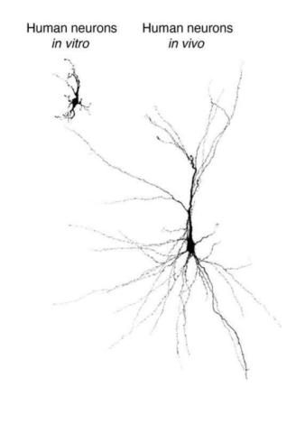
“We’ve been making ever more complicated circuits in a dish using organoids and sophisticated combinations of them, called assembloids. But neurons within these lab dishes are still lagging behind in their development compared with what you’d see in a naturally developing human brain,” Pasca said. Numerous challenges — such as a lack of nutrients and growth factors, blood-vessel-forming endothelial cells or sensory input — hinder development in a lab dish, he said.