Neuroimaging symposium empowers neuroscientists to utilize MRI

Magnetic Resonance Imaging (MRI) is a central technique in neuroscience, enabling researchers to peer deep into the brain to measure neural anatomy and map functional brain networks. But the technique, centered in the physics of electromagnetism and requiring access to a large, expensive MRI scanner, can feel forbidding to researchers without prior exposure to the technology.
The Neuroscience Preclinical Imaging Lab (NPIL) at the Wu Tsai Neurosciences Institute recently welcomed researchers from across Stanford schools and departments to its second annual symposium, geared toward increasing accessibility of brain imaging technologies to scientists throughout campus.

Members of the NPIL community celebrate a successful symposium. Photo by Julia Diaz.
The NPIL — one of Wu Tsai Neuro’s seven community laboratories — provides state-of-the-art MRI technology and expertise to the Stanford community to support preclinical imaging in a wide range of animal research.
The NPIL’s “MRI for Neuroscientists” symposium brought together a diverse audience, from neuroimaging novices to experts in MRI physics, to share firsthand insights about how MRI technologies can advance neuroscience research. At the end of the symposium, the NPIL awarded its second annual Neuroimaging Pilot Grants, which enable scientists to try out new ideas utilizing the cutting-edge facility — whether using MRI for the first time in their research or tinkering with innovative new MRI techniques.
“We encourage neuroscientists across Stanford who haven’t used MRI before to start using the Neurosciences Preclinical Imaging Lab. It is a great resource for scientists interested in applying our technologies to their research,” said lab director, Jieun Kim, PhD.

Bruker manufacturer's rep, Saaussan Madi, explains the specifications of NPIL’s MRI technology. Photo by Julia Diaz.
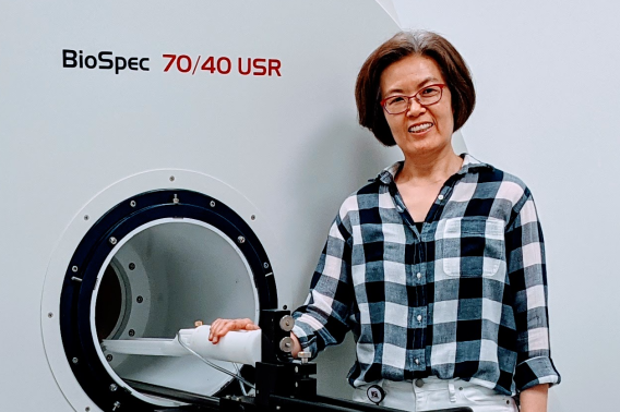
At the symposium, Saaussan Madi, PhD, a senior applications scientist from Bruker, who co-sponsored the symposium, shared the latest advances in MRI technology to give the audience a deeper understanding of the NPIL’s unique 7-Tesla magnet. The magnet is one of only two of its kind in the world because of the combination of its strong magnetic field (which enables higher-resolution imaging) paired with its extra large 40cm bore, allowing the NPIL to support neuroscience studies in a wide range of animal models, from rodents to non-human primates.
Distinguished panelists, including top preclinical imaging scientists from Bruker and neuroimaging experts from Stanford and the National Institutes of Health, shed light on how MRI can elevate scientific discovery, emphasizing the community lab’s role as a resource for researchers across Stanford. Additionally, the discussion explored data management strategies and best practices.

Panelists (l-r) Saaussan Madi, David Leopold, Jieun Kim, and Doug Kelley discuss the many ways scientists can use MRI to advance fundamental neuroscience research. Photo by Julia Diaz.
In his presentation, keynote speaker David Leopold, PhD, chief of the Section on Cognitive Neurophysiology and Imaging and director of the Laboratory of Neuropsychology at the National Institute of Mental Health, shared new perspectives on brain organization made possible by the advent of high-resolution, single-unit fMRI mapping — which allows researchers to visualize individual cells and record activity from single neurons with MRI.
“We want to emphasize that MRI should no longer be considered a low-resolution technique,” said Jin Hyung Lee, PhD, an associate professor of neurology, neurosurgery, and bioengineering and the NPIL’s faculty director.
Five of the nine recipients of last year’s 2022 NPIL Pilot Grant presented the progress of their projects, showcasing the profound impact of MRI technology on their research.
“I am an example of a brand new user. I do not have a background working with MRI, so my project could not have been done without help from the Neuroscience Preclinical Imaging Laboratory,” said Cholawat Pacharinsak, DVM, PhD, associate professor and director of anesthesia, pain management, and surgery at Stanford's Department of Comparative Medicine.
Pacharinsak’s project focuses on understanding the effects of hypothermia on cardiac function, investigating the impact of anesthesia-induced hypothermia on mice. By utilizing MRI techniques, his team aims to assess the severity of cardiac impairment caused by anesthesia.
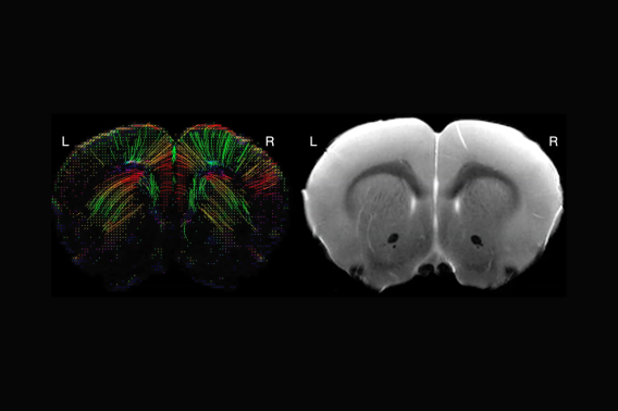
In contrast, Gustavo Chau, a Stanford Interdisciplinary Graduate Fellow and member of the Center for Mind, Brain Computation, and Technology (MBCT) at Wu Tsai Neuro, is an expert in MRI and utilized the NPIL and the pilot grant to further his research. Chau studies white matter microstructural changes associated with absence seizures, characterized by brief lapses in awareness and cognitive deficits. He is developing new techniques on how variations in myelin (the protective fatty layer that covers neurons) affect absence seizures.
“Recent hypotheses suggest that maladaptive alterations in myelination and its interplay with absence seizures may trigger these impairments,” said Chau.

NPIL director Jieun Kim presents the facility's unique 7 tesla magnet. Photo by Nick Weiler.
Attendees and participants networked over a poster session at the conclusion of the symposium. Accompanied by food and beverages, scientists shared their excitement about applying the knowledge gained during the symposium to their ongoing research endeavors.
With an inspiring conclusion, Kim engaged attendees with friendly encouragement to come by the NPIL to talk about their research along with a tour of the facility.
“Never hesitate to reach out to me if you’re interested in using the MRI for your research,” she said.
Learn more about the Neuroscience Preclinical Imaging Laboratory.
2023 Neuroimaging Pilot Grant Awardees
The 2023 NPIL Pilot Grant projects are led by scientists from various departments in Stanford’s Schools of Medicine, and Engineering, including Pathology, Materials Science and Engineering, Neurosurgery, Radiology, Neurosurgery, and Medicine.
Establishing brain morphology and circuit connectivity in rodent models with gene copy number variations of alpha-synuclein
Birgitt Schuele
Pathology, SOM
Harnessing MRI technologies to optimize ventricle imaging in mouse pups
Ryann Fame
Neurosurgery, SOM
Developing a translational diffusion MRI-based biomarker for the therapeutic action of psychedelics
Sedona Noel Ewbank & Alex Ronald Hard
PI: Raag Dar Airan
Radiology, SOM
Investigating Mechanisms of Radio-Resistance in Skull Base Chordoma
Christine K Lee, Jonathan Lamano & Thomas Johnstone
PI: Juan C Fernandez-Miranda
Neurosurgery, SOM
Characterizing a novel mouse model of ALG13 p.N107 using neuroimaging
Juliet Knowles, Richard Coca & Raquel Alvarez
PI: Matthew Wheeler
Medicine, SOM
Neuroimaging of Brain Inflammation Induced by Spinal Cord Injury
Theo Palmer & Vanessa Doulames
PI: Sarah C. Heilshorn
Materials Science & Engineering, SOE

