Event Details:

High-speed imaging of whole-brain activity

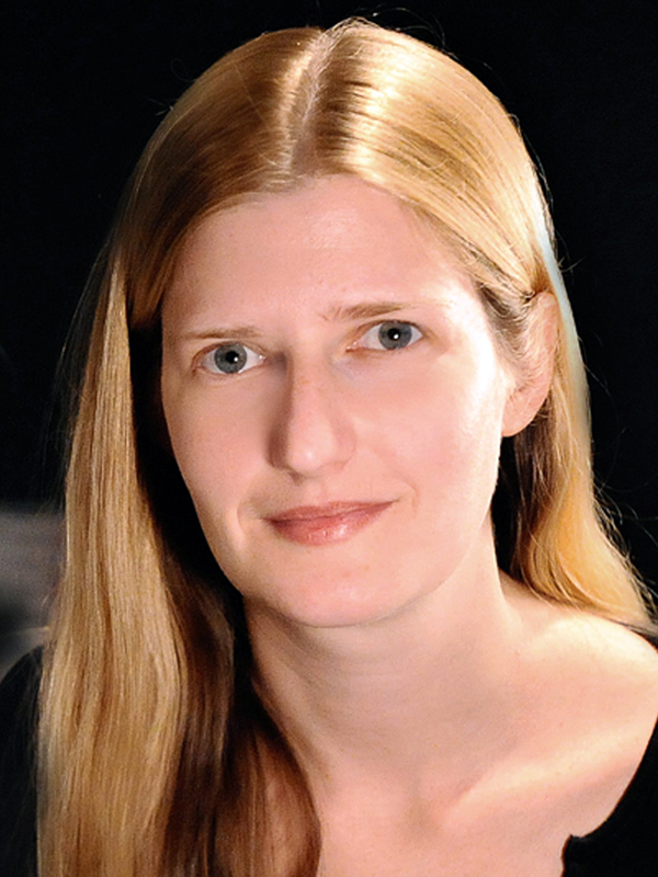
Elizabeth Hillman, PhD
Professor of Biomedical Engineering
Columbia University
Host: Audrey Fan (Zaharchuk Lab)
Abstract
Despite dramatic improvements in in-vivo optical reporters and modulators of neural activity, imaging challenges still limit our ability to capture the activity of thousands of neurons, across large brain regions in awake behaving organisms. My lab has contributed several imaging techniques to address these challenges including swept confocally aligned planar excitation (SCAPE) microscopy for high-speed 3D microscopy and wide-field optical mapping (WFOM) for imaging the whole dorsal cortex in awake, behaving mice.
SCAPE is a high-speed form of light sheet microscopy that can image structure and function in a wide range of living, intact organisms. Unlike most light-sheet systems, SCAPE uses only a single, stationary objective lens at the sample and captures volumes by laterally scanning an oblique light sheet. SCAPE’s features combine to enable very fast (up to 100 volumes per second) imaging in addition to providing high signal to noise with low photobleaching. We are applying SCAPE to imaging awake, behaving organisms such as the freely crawling Drosophila larva, the whole brains of behaving adult Drosophila, zebrafish brain and heart, C. Elegans worms as well as in living mouse olfactory epithelium and the awake mouse cortex. We have also developed numerous technological extensions of SCAPE including systems with much higher spatial resolution, with larger ‘meso’ fields of view, two-photon implementations and systems optimized for high-speed imaging of cleared tissues and for histopathology.
Wide-field optical mapping (WFOM) enables high-speed imaging of both neural activity and brain hemodynamics over the entire dorsal cortical surface in awake, behaving mice. This technique provides very high throughput observations of real-time, bilateral brain activity, providing a way to analyze the interplay between spontaneous behavior and brain-wide neural representations. This platform also enables detailed, longitudinal analysis of changes in neurovascular coupling, brain function and state induced by behavior, drugs and diseases.
Both of these techniques are providing new high-speed, real time views of brain-wide activity in awake, behaving animals. I will present our latest progress on high-speed imaging technique development, and showcase work applying these techniques to understand whole-brain activity in the context of awake behavior.
Related papers
[1] Ma Y, Shaik MA, Kozberg MG, Kim SH, Portes JP, Timerman D, Hillman EMC (2016) Resting-state hemodynamics are spatiotemporally coupled to synchronized and symmetric neural activity in excitatory neurons. Proc Natl Acad Sci U S A 113:E8463-E8471. DOI: 10.1073/pnas.1525369113
[2] Bouchard MB, Voleti V, Mendes CS, Lacefield C, Grueber WB, Mann RS, Bruno RM, Hillman EM (2015) Swept confocally-aligned planar excitation (SCAPE) microscopy for high speed volumetric imaging of behaving organisms. Nature photonics 9:113-119. DOI: 10.1038/nphoton.2014.323