Stanford psychologists explore brain development in facial recognition and reading

By Vedika Kanchan
As children transition from adolescence to adulthood, their brains can grow electrical insulation that supports reading and facial recognition, according to research from the Stanford Psychology Department.
Brain development research has long held that the cortex, the outer layer of the brain, is thicker in children than in adults. Kalanit Grill-Spector, psychology professor and lead author of the study, found that the cortex does not shrink during adulthood as much as initially recorded. Instead, the reason for this difference in thickness is, instead, myelination.
Myelin is an insulating layer in the nervous system that allows quick transmission of electrical impulses along the nerve cells. Grill-Spector and her colleagues propose that the cortex is simply more myelinated in children than adults, meaning that nerve fibers in the brain are more coated in the insulating tissue. Growth of myelin makes it difficult to perceive cortex thickness in MRI scans. Ultimately, this growth also enhances electrical impulses and can make the human brain more efficient, according to Grill-Spector’s research.
New avenues
These findings illuminate the gaps in our knowledge of the human brain. Grill-Spector suggests that her team’s ongoing research and findings will continue to spark more questions about the human brain such as how it develops through infancy.
“We really know very little,” Grill-Spector told The Daily. “It’s actually striking how little we know.”
She suggests that this lack of knowledge partially stems from an inability to use human brains for studies, which she frames as one of the most gaping deficits in brain development research. It is only with new myelin sensitive technology that researchers are better able to scan human brains for cortex thickness.
While researchers seek to understand the human brain, they obtain most of their data from the brains of other primates, like the macaque monkey. The parallels between the brain of a human and the brain of a macaque remain integral to the study of brain development. However, there are clear underlying differences between the brains that are studied and the human brains that researchers seek to apply their learning to, according to Grill-Spector.
Grill-Spector’s research evades the conventional problem that comes with generalizing non-human data to humans. The team examined the brains of 27 children, aged five to 12 years old, and 30 adults. With the use of three magnetic resonance imaging brain scans, researchers were able to find tissue growth in regions linked to facial recognition.
“I couldn’t have done this study ten years ago,” said Grill-Spector. “As technology develops, we have better tools to ask questions.”
The development of new scanning techniques, sensitive to myelin, has found that this tissue growth is linked to myelination. This finding questions the long-standing idea that the cortex thins in its transition from childhood to adulthood.
Breaking grounds
Still, Grill-Spector’s findings offer a small part of the answer. With the Wu Tsai Neuroscience Institute, a Stanford research community dedicated to the study of the brain, researchers are now looking at the same questions in infants, a previously understudied aspect of brain development. The new project examines the human brain, specifically the visual cortex, in the first six months of life.
“So far, we don’t know a lot about how visual cortex develops during that time so there is not a lot of data available,” wrote Mona Rosenke, a Ph.D. student in psychology who worked with Grill-Spector, in an email to The Daily. “Recent advancements in MRI measurement techniques allow us to track anatomical development over time.”
Despite having limited data that explores development in an infant brain, Rosenke suggests that the first six months are incredibly crucial. In this time, infants learn to explore the visual environment around them and recognize faces
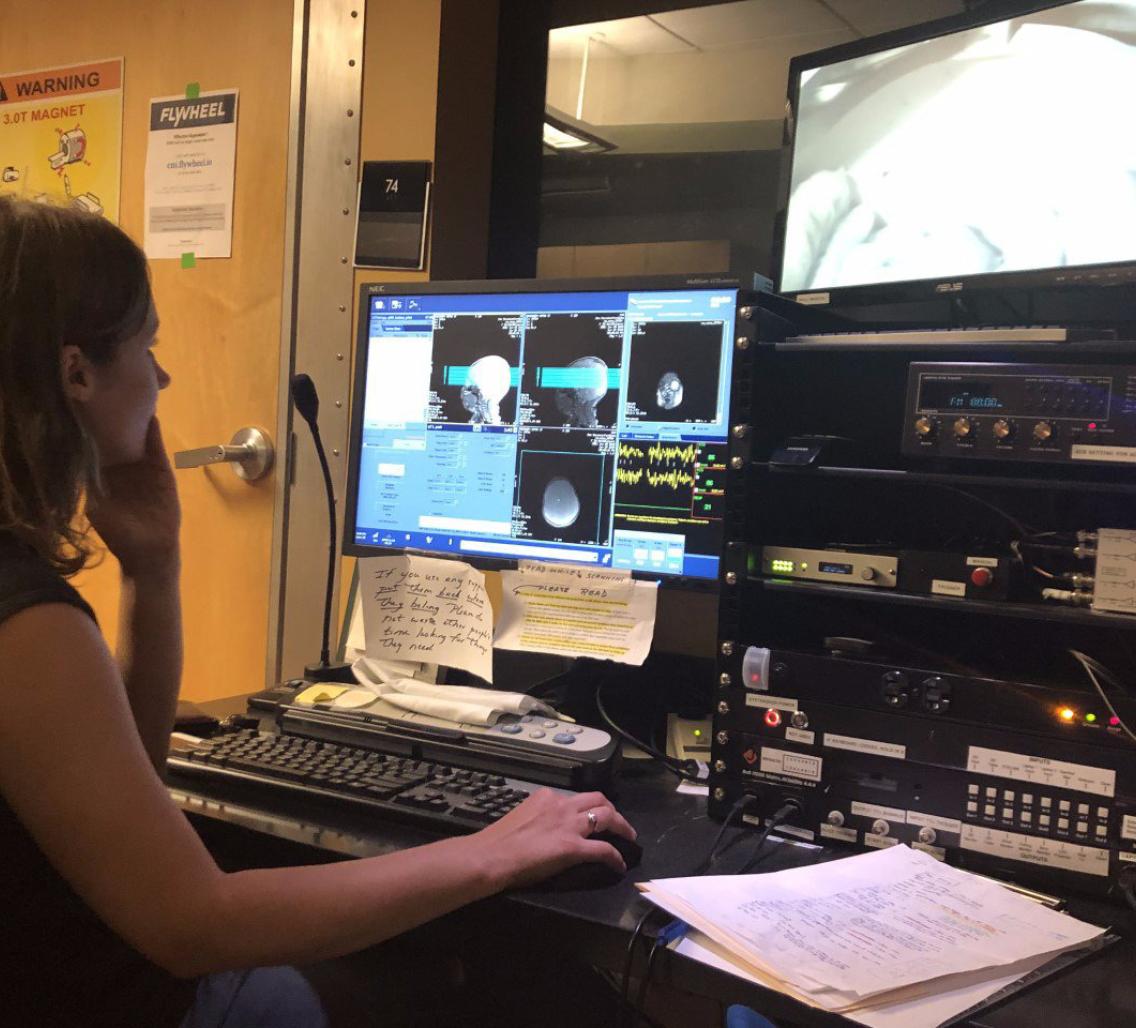
In order to confirm this, the team invites the infant and their parents three times: at less than five weeks old, three months old and six months old. After the parent puts the infant to sleep in an MRI room, researchers use a series of scans to examine where the cells are, changes in tissue properties in the cortex and the connections between different parts of the brain.
It takes a village
According to Rosenke, cross-departmental collaboration was especially crucial to bridging these research gaps. Although this study rests on an intersection of neuroscience and psychology, it is equally rooted in the efforts of MR physics experts that supported the development of sequences that can best measure infant brains and diffusion imaging experts that helped optimize understanding of the connections between brain areas.
Rosenke suggested these learnings were further supported by experts in electroencephalography and computational neuroscience departments who complete the cohesive, but diverse unit of researchers paramount to this study.
Nevertheless, Rosenke says that at the heart of the infant study, is the infants.
“As exciting as this project is, it comes with technical challenges that range from MR sequence development for baby brains to prior experience working with infants,” Rosenke wrote. “Baby brains are so different from those of children and adults.”
With the help of Dr. Ian Gotlib, the Director of the Stanford Neurodevelopment, Affect and Psychopathology (SNAP) Laboratory and his research assistants, the team has been able to work around some of the logistical puzzles of examining infants.
By exploring the brain at its very early stages of development, this collaboration bridges a sobering rift in our understanding of neuroscience. Grill-Spector claims that, for the first time, researchers have had the chance to understand the ties between physical changes in the brain and functional changes in reading and facial recognition.
Contact Vedika Kanchan at vedikak ‘at’ stanford.edu.