Symposium highlights new imaging facility technology and services
Magnetic resonance imaging (MRI) plays a central role in the quest to understand the brain, but researchers not experienced with the technique may find the technology and its physics intimidating.
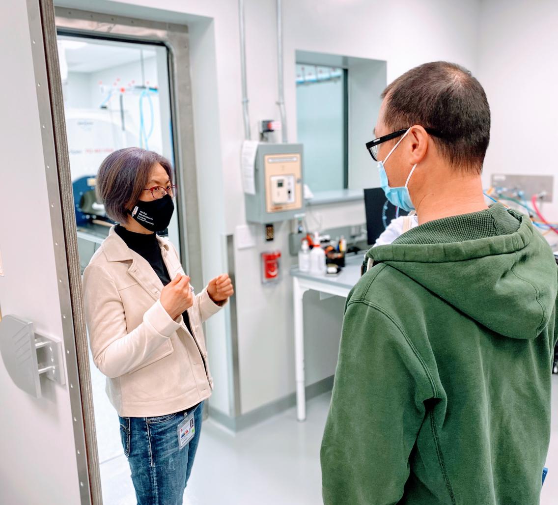
The Neuroscience Preclinical Imaging Laboratory (NPIL) at the Wu Tsai Neurosciences Institute aims to make brain imaging more accessible for researchers of all backgrounds, especially those without prior imaging experience.
At the Inaugural NPIL Symposium on Apr. 15, held in the new Stanford Neurosciences Building, speakers and panelists ranged from expert MRI application scientists to researchers who had just started to explore imaging techniques. Presentations discussed Stanford’s newest MRI imaging facility's potential and explored a variety of MRI research, creating an encouraging environment for new users. At the conclusion of the half-day event, the lab also announced the winners of its inaugural Neuroimaging Pilot Grants, designed to promote experimentation by new users of the magnet.
Early in the day’s sessions, Kristin Granlund, a representative of MRI manufacturer Bruker, outlined the latest advances in the technology, sharing with a stunned audience that with new developments in the technology, scanners may soon be able to image with enough resolution to parse individual cells.
“Part of the goal of this lab is to defeat the notion that MRI is not a high resolution technique,” commented Jin Hyung Lee, associate professor of neurology, neurosurgery and of bioengineering who played a pivotal role in the creation of the new facility and now serves as its faculty director.
Affectionately nicknamed Günther, the NPIL’s 7 Tesla Bruker MRI magnet with a unique 40-cm diameter bore arrived on campus allows for high resolution live imaging of rodents and non-primate animals. The magnet arrived on campus in dramatic fashion in early 2021 and became the first system of its kind to be installed in the United States.
After Granlund’s introduction to the technology, the symposium moved into an “MRI for dummies” panel designed to answer questions researchers might have about the technique but had never had a chance to ask.
“Who here considers themselves an MRI dummy?” asked Lee. Most attendees raised their hands.
“Good,” said Lee, “We specifically made this panel for you.”
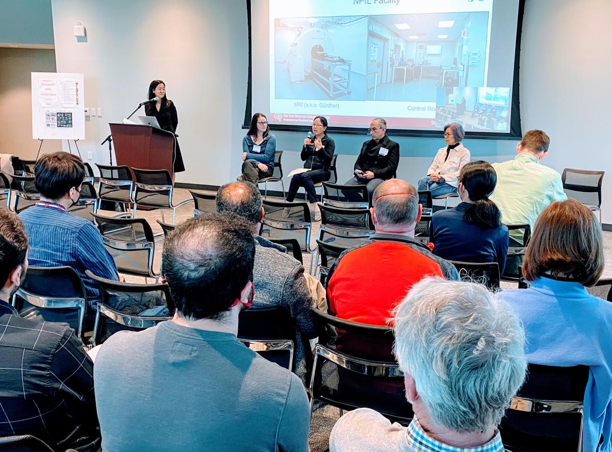
An 'MRI for Dummies' panel demystifies the brain imaging technology at the 2022 Neuroscience Preclinical Imaging Laboratory Symposium. Photo credit Tim Doyle.
The facility itself is also built to be friendly for new users. Five years ago, researchers would have had to have the physics and computing know-how to program their MRI imaging protocols themselves in order to get desired results. In the NPIL, the varying levels of service provided by laboratory director Jieun Kim mean that users can bypass the most complicated steps when working with Günther.
In fact, researchers can choose from three levels of support, Kim explained during the panel discussion. She is available to offer basic guidance on translating research questions into MRI experiments and to complete all the scanning herself. Other researchers familiar with MRI experimental design can request her assistance for operating the scanner only with an approved protocol. Expert users, after training, can use and program the machine themselves to fit their own needs.
“It’s an à la carte service,” said Kim. During the panel, she answered questions without using complex imaging jargon, while also managing to inject doses of humor.
The preclinical imaging lab not only provides instrument support, it also encourages users with unique imaging proposals by offering financial awards. The lab’s Neuroimaging Pilot Grants aim to encourage users of all experience levels to explore imaging in their research. The grants offer up to $5000 to be used for preclinical imaging services. Following the panel, experts and novice users, including three of the first pilot grantees, spoke about how they plan to use MRI in their research.
Sui Wang, assistant professor in ophthalmology, studies retinal development and diseases with rodents. Wang cited imaging the entire shape of the eyeball and the fluid circulation in the eye as the most important functionalities for her research, and the new machine allows her to do both on a longitudinal scale with live animals.
Michael Zeineh, professor of radiology, aims to image the brain before and after a traumatic brain injury. His research will involve imaging pig brains, since their brain structure is similar to that of humans. Through MRI scans, Zeineh hopes to find biological patterns that can inform our knowledge about human brains under traumatic injury.
Tom Sudhof, professor of molecular and cellular physiology and of neurosurgery, conducts research on how molecules affect the formation of synapses. With the MRI, he hopes to connect synaptic action on the molecular level with the imaged activity of neuronal circuits in the brain.
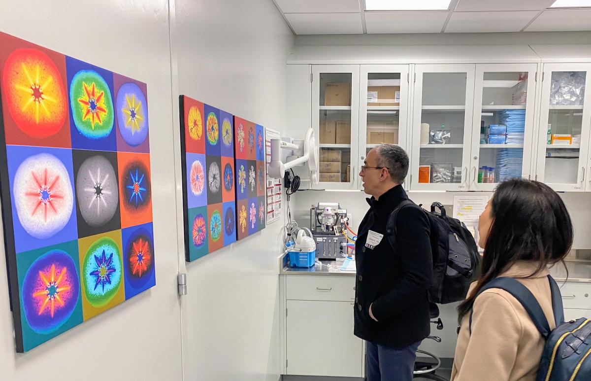
NPIL Symposium attendees got a chance to tour the new MRI facility. Photo credit Tim Doyle.
After the symposium, attendees also had the chance to tour the MRI facility, located in the basement of the Neuroscience Building. While the machine is currently available for use most of the time, Kim expects the schedule to fill up soon, as the campus returns to full capacity.
“If you’re interested in using the MRI facility for your research, reach out to me as soon as possible!”
2022 Neuroimaging Pilot Grant Awardees
Azetidine-Induced Oligodendrogliopathy in Mice
Megan Ann Albertelli & Raymond Sobel
Imaging atrophy and blood-brain barrier function in chronic stroke
Marion Buckwalter
Hypothermia Alteration of Cardiac Function under Isoflurane Anesthesia in Mice
Cholawat Pacharinsak & Monika Kristine Huss
Identification of a behavioral biomarker of concussion
Kyle Lyman & Ivan Soltesz
Magnetic Resonance Imaging of Absence Epileptogenesis
Jennifer McNab
Investigating immune-modulatory effects of luciferase expression in novel syngeneic mouse models of brain cancer
Claudia Petritsch
Unbiased, Brain-wide Analysis of Brain Structure and Neurovascular Integrity Following Deletion of a Cell-Cell Adhesion Module in Astrocytes
Justin Trotter, Alessandra Sclip & Thomas Sudhof
Application of MRI for Studying Primary Congenital Glaucoma
Sui Wang
Neuroimaging of Mild TBI in Pigs
Michael Zeineh, David Benjamin Camarillo & Marios Georgiadis


