‘Kirigami’ electrodes unfold new horizons for brain organoid research
It all started at Peet’s. Ten years ago, neuroscientist Sergiu Pasca was just getting his lab up and running. He had not yet published the research for which he would become renowned: a novel technique for growing 3-D clusters of human brain cells, often called brain organoids, in Petri dishes. Already, Pasca was confident that these organoids and more complex “assembloids”, which mimic many aspects of the growth and maturation of human brain circuits, would soon let scientists study brain development more precisely than ever before, enabling new insights into the early drivers of conditions like autism or epilepsy.
Realizing that potential, however, was a daunting challenge.
“As we began building neural tissues that were ever-more complex, the main limitation was not how we make them, but actually how we’re going to precisely probe and manipulate them,” said Pasca, now the Kenneth T. Norris, Jr. II Professor of Psychiatry and Behavioral Sciences.
That’s when he ran into chemist Bianxiao Cui at a cafe at the James H Clark Center, which sits between the Stanford School of Medicine and the University's chemistry and biology buildings and was then the home of the Wu Tsai Neurosciences Institute. Cui is an expert at designing unusual devices and approaches to record the electrical activity of neurons and other cells. They sat down together, and Pasca told Cui about his as-yet-unpublished organoid research.
“That was amazing and quite eye-opening,” said Cui, the Job and Gertrud Tamaki Professor in the Department of Chemistry. The problem Pasca presented was intriguing: how to record from free-floating clusters of neurons over their months-long development, without catastrophically disrupting their structure and electrical connections.
Solving that problem took over a decade of concerted effort, but Cui, Pasca, and their team have finally cracked it. Their approach—inspired by kirigami, an origami-like art form that involves cutting and folding paper to make intricate sculptures—was reported on January 22, 2024, in the journal Nature Biotechnology. The study was spearheaded by Wu Tsai Neuro Interdisciplinary Postdoctoral Scholar Xiao Yang and Cui lab postdoc Csaba Forro.
Those years of effort, and their ultimate payoff, would have been impossible without the impressive array of expertise housed within a half-mile radius at Stanford, Pasca says—the sort of place where a chemist and a physician-scientist (Pasca trained as a psychiatrist) can meet by chance at a campus cafe and begin a decade-long collaboration—as well as the support of forward-thinking and risk-tolerant organizations like the Wu Tsai Neurosciences Institute.
The challenges ahead had been clear from Pasca and Cui’s first conversation. Traditional recording techniques are designed to measure brain activity in living animals or in cells cultured on a fixed surface. Brain organoids, however, float freely in fluid and are easily damaged by recording devices. And since organoids are much smaller than an entire brain, even a little damage can be catastrophic. Some scientists record from organoids by slicing them in half and placing the cut edge on an electrically conductive surface, but this approach has downsides, too. “In just a few days, they become like a pancake,” Cui said. “They’re no longer free-floating, and lose some of their unique properties.”
Because brain organoids are so different from the tissues neuroscientists have historically worked with, designing approaches for studying and manipulating them demanded a wide diversity of expertise. So Pasca got together several Stanford scientists to found the Stanford Brain Organogenesis Program, which he leads as the Bonnie Uytengsu and Family Endowed Director. With the support of from Wu Tsai Neuro's Big Ideas in Neuroscience program, Pasca, Cui, and their team attacked the problem of recording from organoids with vigor, even having devices fabricated in Europe when clean rooms at Stanford shut down due to the COVID-19 pandemic.
In 2022, Pasca, Cui, and Stanford Brain Organogenesis Program member Zhenan Bao, the K. K. Lee Professor of Chemical Engineering, published a paper reporting on the fruit of their labors: a flexible, mesh-like electrode that could be partly inserted into an organoid.
But their quest wasn’t over. “It did not work well,” Cui says. Because the organoids were only affixed to the electrodes on one end, the fluid flowing around them would sometimes tear them entirely free.
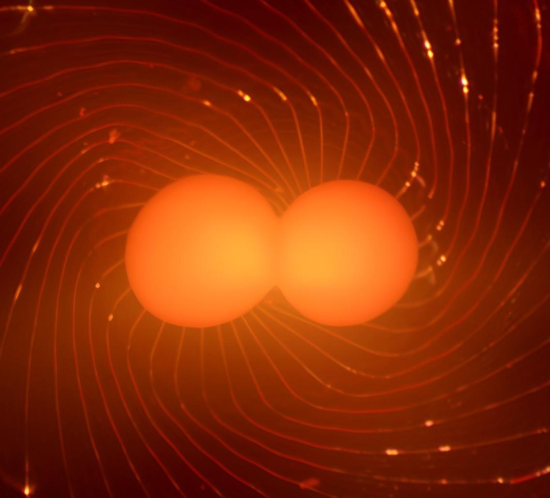
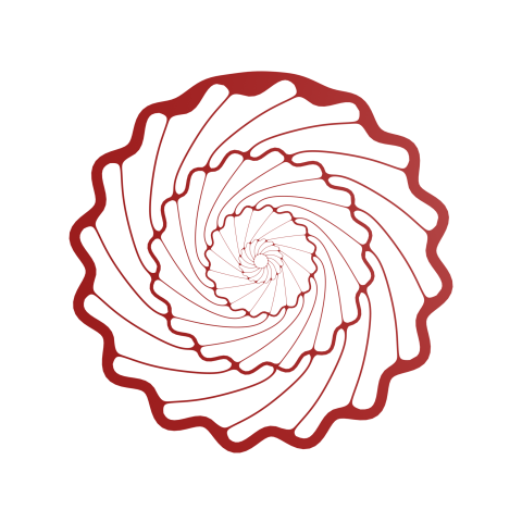
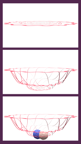
Their next attempt, however, proved far more successful. Instead of plugging the electrode into the organoid, like a stick poked into a marshmallow at a campfire, the team used a kirigami-inspired approach to fabricate a device that could fully cradle the organoid. Initially, the device looks like an intricate, electrically conductive doily; once researchers place an organoid at its center, however, it bends and twists into a basket-like shape. The organoid then continues to grow, eventually engulfing the device’s supportive strands.

For the hammock-like electrode to work, it needed to be flexible enough to allow the organoid to grow naturally and stable enough to stay intact over many months of recording. Balancing those objectives was tricky, so the researchers used computer simulations to aid their iterative design process. The result is extremely effective—and extremely delicate. “It’s easy to tear it,” Cui said. “You touch it with a pipette, you’ll break it.”
Already, though, Pasca, Cui, and their team have demonstrated that the electrode can record from organoid cells for months—orders of magnitude longer than previous approaches— apparently without disturbing its natural development.
This success allowed the team to rigorously answer a question that had previously proved tricky to address: when, exactly, do organoids become electrically active? Because this new device can record from organoids for so long, with no apparent effect on their development, the researchers were able to demonstrate that their organoids started firing action potentials when they were around 100 days old.
“We’ve never been able to continuously monitor one neural organoid to see when electrical activity emerges,” Pasca said. “And that’s actually a fundamental question, even for understanding human brain development.”
The team has their sights set on even more ambitious questions — Pasca’s team has recently grown groups of organoids representing different components of brain tissue—termed “assembloids” that can capture the formation and activity of specific human brain circuits and to study their susceptibility to neurodevelopmental disease.
They envision next-generation kirigami devices that incorporate not only electrodes but also sensors for substances like oxygen and chemical neurotransmitters, or LEDs that can stimulate cells using optogenetics, a common tool in neuroscience research developed by Stanford Brain Organogenesis Program member Karl Deisseroth, the D.H. Chen Professor of bioengineering and of psychiatry.
Like any new methodology, these kirigami electronics will see their fullest realization when taken up by the scientific community, which will be accelerated by the popular hands-on course on neural organoids and assembloids the Brain Organogenesis team hosts each year on the Stanford campus, drawing scientists from around the world.
Above all, Pasca said, it’s key to recognize that this is a tool designed to advance researchers’ ability to understand the mysteries of how the human brain organizes itself early in life — and hopefully develop new approaches to repair defective neural circuits. “I think the power of this technology will be in how people use it.”


