Project Summary
The immune system is subjected to neuroendocrine regulation and control by the brain. One such example is the induction of glucocorticoid (GC) in infectious diseases. GC is synthesized and released by the adrenal glands via the hypothalamic-pituitary-adrenal gland (HPA) axis which is initiated from the hypothalamic paraventricular nucleus (PVN). GC regulates many immune and physiological responses. Persistent high GC levels often result in severe immune suppression and metabolic abnormalities. However, GC dysregulation is induced during an infection remains unclear.
Our recent analysis of the host response to influenza virus infection showed that while mice infected with a sub-lethal dose of the virus all survived, those lacking functional gd T cells, a subset of T cells, all succumbed. These mice have high levels of circulating GC and lowering the GC level with a GC synthesis inhibitor rescues these animals. During the infection, mice lacking functional gd T cells also had severe breathing abnormalities and much reduced blood oxygen levels, indicating severe hypoxia. As the brain is the highest oxygen-consuming organ, and hypoxia alters the GC response, we hypothesize that during an influenza virus infection, mice without functional gd T cells develop severe breathing problems in the lung, leading to hypoxia and GC dysregulation in the brain.
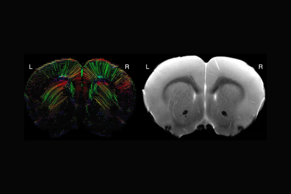
Here, we propose to analyze the brain responses to influenza virus infection with MRI imaging, focusing on the state of hypoxia. It is part of the effort to determine “mechanisms of immune response mediated glucocorticoid dysregulation in influenza virus infection.”
Project Details
Funding Type:
Neuroimaging Pilot Grant
Award Year:
2024
Lead Researcher(s):
